DIGESTIVE SYSTEM
All snakes are carnivorous so the gastrointestinal tract is a relatively simple, linear duct, which extends from the oral cavity to the cloaca (Fig. 5.1).
Dentition
Snakes swallow their prey whole without mastication so the teeth function solely in food prehension.
Consequently, they are long, thin and backwardly curved to prevent the escape of prey. All snakes have pleurodont teeth that are attached to the medial jawbone and are continually being replaced by new teeth lying in reserve in the gums (Fig. 5.18). Each tooth lasts only a few months before being shed and swallowed with the prey. In venomous species some maxillary teeth are modified into fangs (Edmund 1970).The number of teeth varies between species but most snakes seen in veterinary practice have six rows of teeth in total: one row on each lower jaw and two rows on each maxillary and palatine or pterygoid bones of the upper jaw (Edmund 1970) (Figs. 5.19 and 5.20). Copious amounts of saliva are produced from the palatine, lingual, sublingual and labial salivary glands during swallowing, which moistens and lubricates the prey.
Venom glands
These are modified labial salivary glands, that produce venom that immobilize the prey preventing damage to the delicate skull. The venom contains collagenases, phospholipases, proteases and injection into the prey is under voluntary control (Bellairs 1969c).
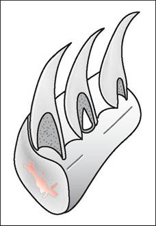
Figure 5.18 • All snakes have pleurodont teeth. These are thin and backwardly pointing to prevent escape of prey.
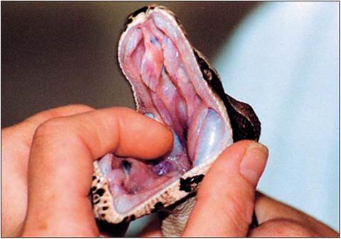
Figure 5.19 • Ventral view of maxillary arcade. Most species seen in practice have four rows of upper teeth. (Photo by Janet Saad)
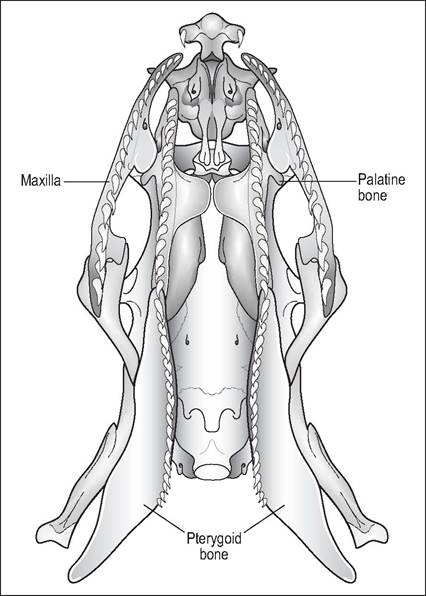
Figure 5.20 • Ventral view of maxilla showing maxillary and palatine/ pterygoid dental arcades.
Rear-fanged (opisthoglyphous) snakes
In about one third of the colubrids the caudal labial gland becomes modified into a distinct capsular gland lying behind the eye and just above the lips. This gland is known as Duvernoy's gland and its function is to secrete venom to immobilize prey. Venom passes from this gland into a modified tooth at the caudal maxilla. These rear fangs are grooved and able to inject venom into prey. Like all teeth the fangs are shed regularly to be replaced by the reserve fangs (Bellairs 1969c; Evans 1986; Pough 1998a, 1998b).
In general the back-fanged snakes are not so venomous, the exception being the Boomslang (Dispholidus typus), which can cause fatalities (Fig. 5.21). The main aim of the venom is actually to incapacitate the prey so that it cannot damage the mouth while being eaten.
Front-fanged snakes
In these snakes the venom gland is large, separate from the labial glands, and lies behind the eye. A single long duct runs rostrally into fangs situated at the rostral maxilla. Some cobras can actually spit venom over 2 m away. In Elapidae the fangs remain erect and cannot fold (proteroglyphous) (Pough et al. 2002).
Viperidae have even more highly modified fangs (soleno- glyphous). They are so long that when the mouth is closed the fangs lie folded backwards in a sheath along the roof of the mouth in an area of no teeth (diastema). The shortened maxilla is hinged and mobile. When the mouth opens the pterygoid muscles contract, pulling up the palatopterygoid so that the fangs are raised for striking (Pough et al. 2002) (Fig. 5.22).
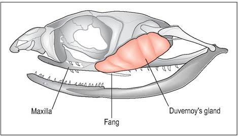
Figure 5.21 • Rear fangs - Boomslang (Disphofidus typus) showing location of Duvernoy’s gland and position of rear-grooved fangs.
Tongue
The tongue is long, slender and forked and lies in a sheath beneath the glottis and rostral trachea (Fig. 5.23). It is very mobile and can be protruded through the lingual notch or fossa without the snake opening its mouth.
It functions in olfaction, taste and touch (Fig. 5.24).Gastrointestinal tract
The esophagus is relatively thin walled and amuscular as the axial musculature plays a major role in the transportation of
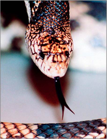
Figure 5.23 • Forked tongue of snake. (Photo by Janet Saad)
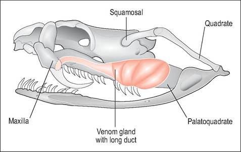
Figure 5.22 • Front fangs - Rattlesnake (Crotafus sp.) showing location of venom gland with duct opening into grooved front fangs. The fangs are folded but the hinged maxilla can be raised to erect fangs for striking.
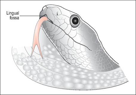
Figure 5.24 • The tongue is very mobile and can be protruded through the lingual fossa without the snake opening its mouth.
food to the stomach. It is highly distensible to allow for prey, which may remain alive there for many hours. Often the only distinguishing feature between the stomach and esophagus is that the stomach has a more glandular mucosa. The stomach is fusiform and there is no well-defined cardiac sphincter, causing easy regurgitation of food (Figs. 5.1 and 5.4).
The esophagus also plays a role in food storage because the stomach is relatively small and may not be able to accommodate the entire prey (particularly with cannibalistic species which often consume prey as long as themselves). Digestion begins as soon as even part of the prey reaches the stomach and is a rapid process. Absorption however is very slow. As the whole prey is utilized, including the skeleton, it may take up to 5 days for a large snake to digest a rat. Only the keratinous structures like fur are finally excreted as an undigested pad called the felt (Fig. 5.25).
GENERAL INTEREST
In egg-eating snakes (Dasypeltis spp.) the cranial esophagus is closely attached to the first 30 or so presacral vertebrae. These vertebrae have modified ventral spines against which the shell is crushed by longitudinal bands of muscle.
The egg contents are expelled into the stomach while the broken shell is regurgitated up the esophagus.CLINICAL NOTE
Figure 5.25 • Bull snake with esophageal and gastric impaction after the owner changed the diet from mice to rats. The combination of lack of humidity and increased size and fur length of prey contributed to a fatal impaction.
When stomach-tubing snakes, infuse slowly and hold them vertically for 30 seconds post feeding to prevent regurgitation through the weak cardiac sphincter.
The liver is elongated and may be divided into two to three separate lobes. As snakes consume large meals infrequently a gall bladder is essential to help digest fat. The pancreas is ovoid and found caudal to the gall bladder on the mesenteric border of the duodenum (Fig. 5.1). In some species the spleen is adherent to the pancreas, creating the spleno- pancreas. The small intestine is fairly straight and a cecum is present is some Boidae species.
The large intestine is separated from the cloaca by a distinct fold. Paired fat bodies, which are often vascularized, lie in the caudal celomic cavity. In snakes the cloaca is linear rather than round and is divided into three sections by mucosal folds. Cloacal scent glands are present in some snakes and serve as a warning mechanism by producing foul smelling secretions (Evans 1986).