RESPIRATORY SYSTEM
Upper respiratory tract
The glottis lies forward and is easily visualized making intubation for anesthesia very easy. It is very mobile and can be extended laterally while feeding to allow breathing while ingesting prey (Fig.
5.17). It lies against the choana dorsally when the mouth is closed. The trachea has incomplete car-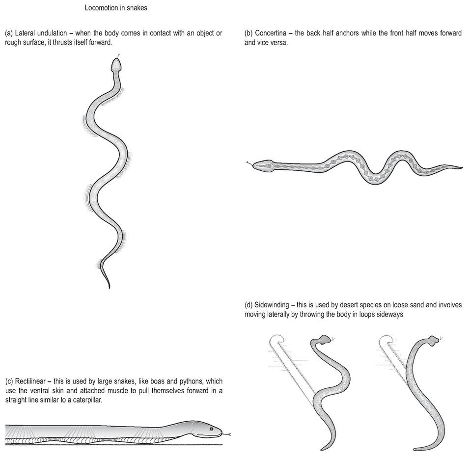
Figure 5.14 • Locomotion in snakes.
(a) Lateral undulation - when the body comes in contact with an object or rough surface it thrusts itself forward
(b) Concertina - the back half anchors while the front half moves forward and vice versa
(c) Rectilinear - This is used by large snakes, like boas and pythons, which use the ventral skin and attached muscle to pull themselves forward in a straight line similar to a caterpillar
(d) Sidewinding - this is used by desert species on loose sand and involves moving laterally by throwing the body in loops sideways
tilaginous rings, with rigid cartilage ventrally and the dorsal fourth being membranous (Funk 1996). Like all reptiles, snakes have a poorly developed mucociliary apparatus and rely on body positioning to help clear mucus and inflammatory exudates. They have no vocal cords but hiss by forcing air through the glottis - the pitch will depend on the width of the aperture and the loudest noise is made during expiration (Bellairs 1969d; Liem et al. 2001b).
CLINICAL NOTE
Snakes have no effective cough reflex so it is possible for experienced handlers to intubate a conscious animal and induce anesthesia by isofluorane. However, this inability to cough up exudates make the snake very susceptible to pneumonia.
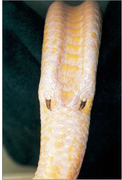
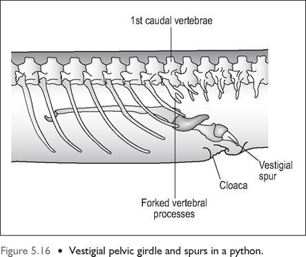
Figure 5.15 • Albino male Burmese python (Python molorus) showing vestigial spurs.
the right lung is slightly longer (Evans 1986; McCracken 1999). The right lung extends from the heart to just cranial to the right kidney (Figs. 5.1 and 5.4).
Structurally, the cranial lung is simple and unicameral (single chambered), with a good blood supply, and performs the air exchange. Like in the Scincidae lizards, the caudal third is non-respiratory and functions like an airsac (Perry 1989). Aquatic snakes have an airsac that extends caudally to the cloaca and acts as a buoyancy aid (Bellairs 1969d).
In some snakes the vascular portion of the lungs extends into the dorsal trachea, creating a saccular extension to the tracheal rings that is capable of gaseous exchange (McCracken 1999).
CLINICAL NOTE
Snakes have a very fragile lung so perform intermittent positive-pressure ventilation (IPPV) with care, so as to avoid lung rupture.
The respiratory cycle
Respiration is controlled by the dorsal and ventrolateral sheet of intercostal muscles, which extend along almost the entire length of the trunk. Some snakes also use the avascular airsac like bellows in order to ventilate the lungs when the passage of food compresses them. In snakes inspiration is both passive and active. Relaxation of the expiratory muscles
Lower respiratory tract
In keeping with body elongation many snakes have evolved one functional lung. The viperids have only one lung; the colubrids have one functional lung (the left lung is vestigial) and the more primitive boids have two saccular lungs, although
Figure 5.17 • Open mouth of Rat snake (Elaphe obsoleta) showing open glottis. In snakes the trachea is very mobile and can be extended out of the mouth to allow breathing while swallowing large prey. (Photo by Janet Saad)
starts the passive part of inspiration. The intercostal muscles then contract, decreasing intrapulmonary pressure and resulting in active inspiration. Passive expiration then occurs as these muscles relax and the lung recoils (Wood & Lenfant 1976).