DIGESTIVE SYSTEM
Feeding behavior
Guinea pigs are crepuscular and thus feed at dawn and dusk. They are strict herbivores with molar teeth suited for grinding vegetative matter and, like rabbits, exhibit cecotrophy.
They are also fastidious eaters that learn early in life what to eat.Guinea pigs grab stalks by the base and tear them off by a backward and upward thrust of the head. In the wild much of the guinea pig's water requirements would come through its greens, but in captivity water must be supplied.
CLINICAL NOTE
When supplied with a water bottle guinea pigs do not lick the drops but put their whole mouths around the nozzle, creating a slurry which can easily block it. This can lead to rapid dehydration in guinea pigs fed solely on dry food diets.
Digestive physiology
The normal gastric emptying time is 2 hours and total gastrointestinal transit time averages 20 hours (range 8-30 hours). If cecotrophy is accounted for then total transit time is 66 hours. Cecotrophy is performed 150-200 times daily and is essential for fiber and protein digestion (Ebino 1993). Young guinea pigs initially populate their gut by eating the sow's droppings. The gut flora is mainly gram-positive bacteria and anaerobic lactobacillus, but coliforms, yeasts, and clostridia are also present in small numbers (Cheeke 1987; Harkness & Wagner 1995; Huerkamp et al. 1996).
Guinea pigs digest fiber more efficiently than rabbits (Cheeke 1987). Unlike in rabbit and rats, satiety in guinea pigs is governed by the distension of the gastrointestinal tract as appetite does not increase with added cellulose to the diet (Cheeke 1987; Harkness 1990; Harkness & Wagner 1995). A crude protein level of 18-20% is needed for growth and lactation, and a minimal crude fiber level is 10% (Huerkamp et al. 1996).
Vitamin C
Guinea pigs lack the enzyme L-gulonolactone oxidase, which synthesizes ascorbic acid from glucose.
Ascorbic acid is necessary for the production of hydroxylysine and hydroxyproline, both essential for collagen synthesis in connective tissues. Abnormal collagen results in leaking blood vessels and hemorrhage in the joints, gums, and intestines. Collagen also anchors the teeth in the sockets so hypovitaminosis C can lead to dental problems (Cheeke 1987; Huerkamp et al. 1996; Navia & Hunt 1976).Adult non-breeding guinea pigs need 5 mg/kg of vitamin C daily in their diet (Harkness & Wagner 1995). Young growing animals have the highest demand for vitamin C; scurvy can develop within 2 weeks on a deficient diet.
Metastatic mineralization
The guinea pig lays down soft tissue calcification with relative ease. Why this occurs is unclear but it may be related to dehydration and mineral imbalances. Lesions can be seen in animals over 1 year of age (Huerkamp et al. 1996).
CLINICAL NOTE
When supplementing guinea pigs with vitamin C it is important to avoid any human multivitamin products as these can be too high in Vitamin D and lead to metastatic calcification (Huerkamp et al. 1996).
Dentition
There are 20 teeth and the dental formula is 1/1, 0/0, 1/1, 3/3. All teeth are rootless (aradicular) and constantly growing (Vaughan 1986). As a consequence of this malocclusion can occur in both molars and incisor teeth. The chisel-shaped incisors are white. The maxillary cheek teeth are angled laterally, the lower teeth are arched medially toward the tongue (Breazile & Brown 1976; Cooper & Schiller 1975b).
CLINICAL NOTE
In guinea pigs with molar malocclusion the maxillary cheek teeth overgrow laterally into the buccal mucosa while the mandibular cheek teeth arch medially, causing tongue entrapment and subsequent anorexia.
Oral cavity
The oral cavity is small and there is a large elongated tongue. The upper and lower cheek folds can fold inwards during gnawing, dividing the mouth into two regions. Caudally the mouth communicates with the pharynx and is lubricated by salivary glands.
The tongue is large and elongated and covers most of the floor of the mouth and oropharynx. It is bounded laterally by the mandible. The rostral two thirds lie free and caudally it is raised into a mound where there are extensive papillae (Cooper & Schiller 1975c). The muscles of mastication are well developed, reflecting the gnawing and grinding behavior of this species (Breazile & Brown 1976).
Salivary glands
There are five pairs of salivary glands: parotid, mandibular, zygomatic, major and minor sublingual. The ducts enter the oral cavity near the molar teeth. The two mandibular glands
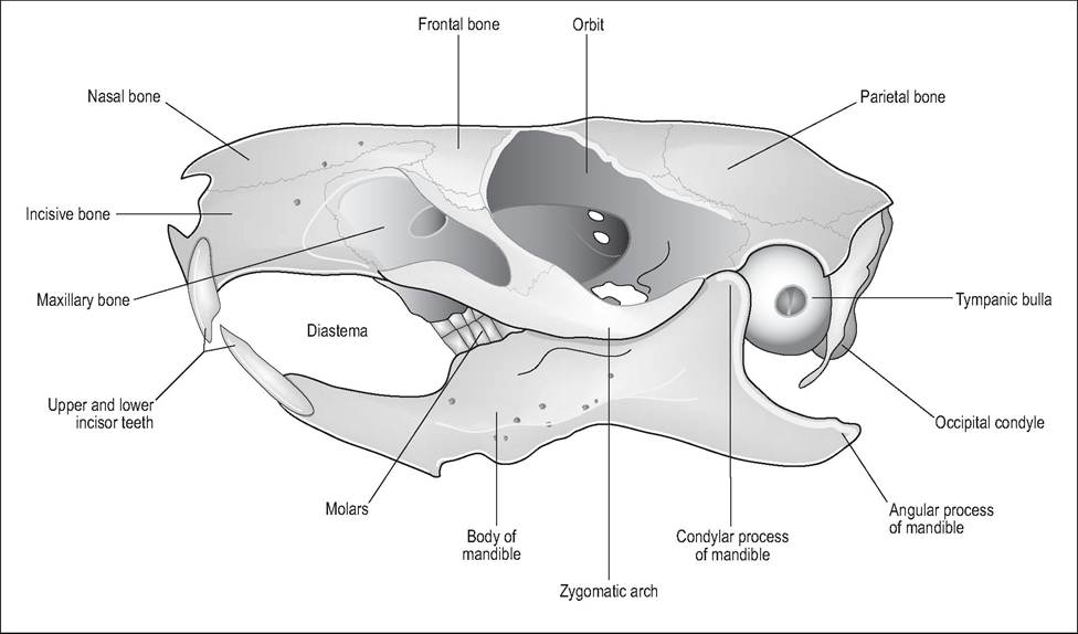
Figure 9.7 • Lateral view of guinea pig skull. From Popesko, P., Rajtova, V., & Horak, J. (1990) A colour atlas of anatomy of small laboratory animals. Vol. I. Aylesbury, UK: Wolfe with permission.
come in contact with each other in the ventral midline (Breazile & Brown 1976; Timm et al. 1987).
Pharynx
The nasopharynx and oropharynx are separated by the soft palate, which communicates with the oral cavity via the palatal ostium (see Respiratory system) (Timm et al. 1987). Guinea pigs do not have tonsils but have lymphoid nodules in folds in the wall of the pharynx.
Esophagus
This runs dorsal to the trachea in the midline and lies to the left as it enters the thoracic inlet. It is lined by stratified squamous epithelium, proximally by striated muscle, and distally by smooth muscle. It enters the cardiac portion of the stomach at an oblique angle near the lesser curvature.
Stomach
Guinea pigs are mongastric but, unlike the rat and hamster, their stomach is completely glandular. The stomach lies in the left cranial portion of the abdomen. The lesser curvature of the stomach is very small and the angle formed by it and the esophagus is called the angular notch. The greater and lesser omentum extend from the stomach, as in other species (Cooper & Schiller 1975c).
Intestines
The small intestine occupies the right side of the abdomen and measures about 125 cm in length. There is little to distinguish the different parts of the intestine. Lymphoid nodules (Peyer patches) are found in the lamina propria. The large intestine begins at the ileocecal valve and terminates at the anus (Breazile & Brown 1976; Cooper & Schiller 1975c).
Cecum
The cecum is a large, thin-walled, green-brown sac filling most of the left ventral abdominal cavity (Fig. 9.9). This is the largest dilation of the alimentary canal, being 15-20 cm long and containing 65% of the gastrointestinal contents. Externally, three white muscular longitudinal bands are visible: the dorsal, ventral and medial teniae coli. These being shorter than the cecum create saccular outpouchings called haustra (Cooper & Schiller 1975c).
Colon
The colon is dark green and 70 cm long. Although it has an ascending, transverse, and descending portion named accord-
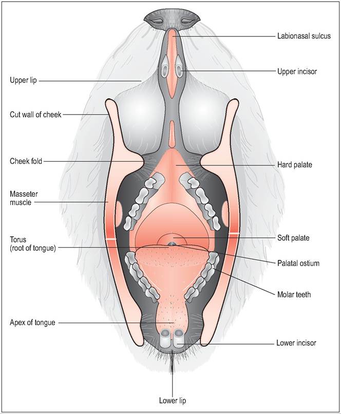
Figure 9.8 • Open mouthed view of guinea pig showing molar teeth and palatal ostium. From Popesko, P., Rajtova, V., & Horak, J. (1990) A colour atlas of anatomy of small laboratory animals. Vol. 1. Aylesbury, UK: Wolfe with permission.
ing to location in the abdominal cavity, it is functionally divided into the proximal (20 cm) and distal colon (50 cm).
The proximal colon plays a role in separating digesta into fecal pellets and cecotrophs. Although there are no haustra outpouchings as in the rabbit, the mesenteric side of the colonic mucosa folds to form a longitudinal furrow. This furrow aids in separating the high protein digesta from the poor quality protein destined to become fecal pellets. In the proximal colon this furrow becomes very deep and is lined by mucous cells which trap bacteria and high protein particles. These are then transported by antiperistalsis back to the cecum for further fermentation (Cheeke 1987; Holtenius & Bjornhag 1985).
Liver
The liver is the largest gland, is red-brown in color, and smooth. There are six lobes: right, medial, left lateral, left medial, caudate, and quadrate. The gall bladder is well developed and lies medially in the quadrate lobe (Breazile & Brown 1976; Cooper & Schiller 1975c).
Pancreas
This is triangular, pink-red and has three lobes. It lies in contact with the descending duodenum.