DIGESTIVE SYSTEM
Rats are omnivores and feed frequently. Food is grasped in the forepaws, chipped by the incisors, and ground down by the molars. Gastrointestinal transit time varies from 12 to 24 hours.
Food restriction, as opposed to ad lib feeding, has been found to increase longevity and decrease incidence of tumors in ageing rats (Koolhaas 1999; Yu 1994).Oral cavity
The tongue is about 30 mm long and 8 mm wide from rostral tip to epiglottis (Fig. 10.13). It is compressed in the middle to allow space for the molar teeth. The dorsal surface has a caudal prominence called the lingual torus, which has abundant gustatory and mechanical papillae (Bivin et al. 1979; Hebel & Stromberg 1986c).
There are no tonsils. The hard palate has prominent ridges and is distinguished from the soft palate by a pale line in the mucous membrane (Bivin et al. 1979). Taste buds are found throughout the oral cavity from the dorsal tongue, along the rostral edge of the soft palate, and on the hard palate.
Dentition
There are 16 teeth and the dental formula is 1/1,0/0,0/0,3/3. The cheek tissue can be drawn into the diastema, closing off the back of the mouth to allow the rat to gnaw on hard substances without having debris pass into the pharynx (Bivin et al. 1979).
Incisors
As is the case with all rodents, the incisive bone is well developed. The incisors erupt about 10 days after birth, the molars on day 19 and all teeth are in wear by 6 weeks. Rats are monophyodont, meaning they only produce one set of teeth (Hebel & Stromberg 1986a; Schour & Massler).
The incisors are open rooted (aradicular) and constantly growing so need to be worn down by gnawing. The incisors grow in the shape of a spiral, the upper incisors being more tightly curved than the lower (Fig. 10.16). The ability of the temperomandibular joint to allow cranial and caudal jaw movement keeps the tips sharp by gnawing.
The lingual side has softer dentine so wears down faster, creating the appearance of the bevel of a hypodermic needle. The incisors have a yellow-orange color due to the presence of iron pigments and this deepens with age (Schour & Massler).CLINICAL NOTE
At rest, the lower incisors lie behind the upper incisors and the length of the lower crown is three times as long as the upper crown. This is normal and should not be mistaken for malocclusion (Fig. 10.2).
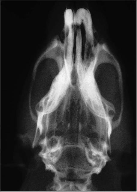
Figure 10.11 • Dorsal radiograph of rat skull.
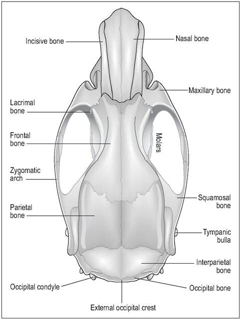
Figure 10.12 • Dorsal view of rat skull.
The rate of eruption is balanced by the rate of attrition so the length of the teeth remains constant. When a tooth is fractured or maloccluded the eruptive forces increase 2-3 times, causing rapid elongation of the tooth. The teeth usually grow in a spiral fashion, often perforating the lips (Schour & Massler).
Molars
These provide the grinding action on the food. They are closed rooted (brachiodontic) and stop growing at about 125 days after birth. The size of the molars decreases from Ml to M3 (Hebel & Stromberg 1986a). The masticatory surface has transversely orientated enamel folds with nine enamel-free cusps arranged in three rows of three. The upper and lower jaws are of equal width but only one side will be in apposition at a time during chewing.
Muscles of mastication
Rats, like all myomorphs, have a well-developed zygomatic arch and strong jaw muscles (Fig. 10.17). The masseter, temporalis, and pterygoideus close the jaw; the digastricus opens the mouth and draws the mandible backward (Hebel & Stromberg 1986b).
The masseter muscle (medial and lateral) is the most powerful and extends from the lateral zygomatic arch to the mandible. In all myomorphs a slip of the medial masseter runs through the infraorbital canal to insert on the muzzle.
This gives a strong cranial pull on the lower jaw and aids grinding and gnawing (King & Custance 1982). The temporalis muscle originates from the temporal fossa and inserts on the coronoid process and medial mandible. The pterygoideus muscle extends from pterygoid/palatine bone and inserts on condyloid and medial angular process. The digastricus muscle, which opens the mouth, arises from the occipital bone and inserts just caudal to the mandibular symphysis (Hebel & Stromberg 1986b).Salivary glands
These consist of the greater salivary glands (parotid, mandibular, and greater sublingual) and the minor salivary glands (sublingual, buccal, palatine, and lingual). The parotid gland is quite diffuse and extends from behind the ear almost to the shoulder (Sharp & LaRegina 1998). The mandibular salivary gland lies in the ventral cervical region about 10 mm rostral to the thoracic inlet (head extended). It borders the mandibular lymph node rostrally. The greater sublingual gland is also tightly attached to this gland rostrally (Bivin et al. 1979; Hebel & Stromberg 1986c).
216
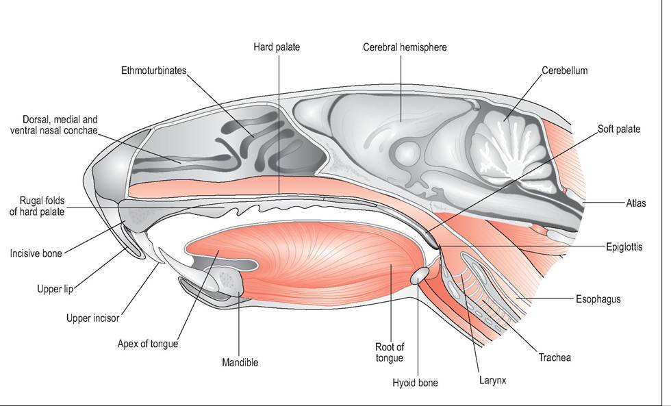
Figure 10.13 • Midsaggital view through head of rat. The larynx is placed high in the oropharynx where it can directly access the nasopharynx, making rats and rodents obligate nose breathers. From Popesko, P., Rajtova, V., & Horak, J. (1990) A colour atlas of anatomy of small laboratory animals. Vol. 2. Aylesbury, UK: Wolfe with permission.
The salivary glands and lymph nodes in the ventral cervical region are covered by extensive areas of brown fat, which extends from the mandible to the axilla and should not be confused with lymph nodes or glands (Greene 1962).
CLINICAL NOTE
Rats can get Sialodacryoadenitis virus (SDAV) which causes inflammation and edema of the cervical salivary glands and lymph nodes, creating the appearance of mumps. SDAV is highly contagious and usually self-limiting, although affected rats pose an anesthetic risk due to enlarged glands pressing on the respiratory tract (Fallon 1996).
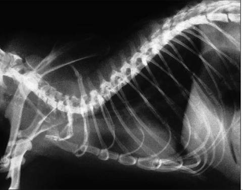
Figure 10.14 • Lateral radiograph of thorax.
Esophagus and stomach
The abdominal muscles and linea alba are very thin. The esophagus runs slightly to the left in the cervical region and then along the dorsal midline. It has a diameter of approximately 2 mm and has skeletal muscle along its length. The esophagus enters the stomach in the middle of the lesser curvature. A limiting ridge at this junction prevents rats from vomiting (Bivin et al. 1979).
The stomach lies transversely, caudal to the rib cage on the left side with its parietal surface covered by the left liver lobe. A lobulated cushion of fat (which is embedded in the mesorchium or mesovarium) is sandwiched between the stomach and the abdominal wall. The oblong spleen is also in contact with the greater curvature.
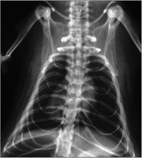
Figure 10.15 • Ventrodorsal view of thorax in rat. Note the well-
developed clavicular brace.
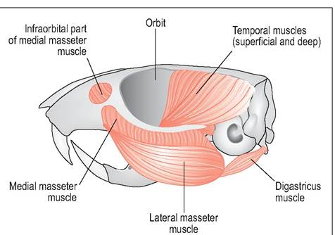
Figure 10.17 • Muscles of mastication in the rat.
Note the powerful masseter muscle (medial and lateral), which extends from the lateral zygomatic arch to the mandible. From Popesko, P., Rajtova, V., & Horak, J. (1990) A colour atlas of anatomy of small laboratory animals. Vol. 2. Aylesbury, UK: Wolfe with permission.
into the cecum lies close to the opening of the colon (Komarek et al 2000)(Figs. 10.19 and 10.20).
Large intestine
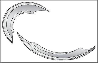
Figure 10.16 • The correct alignment of incisor teeth in the adult rat. The lower incisors lie behind the upper at rest and the ends should be sharp like a chisel.
The comma-shaped cecum commonly lies in the left caudal abdomen, although its long mesentery means its position varies quite considerably (Hebel & Stromberg 1986c; Komarek et al. 2000b). Although it has no septa dividing it, as seen in other rodents, it can be divided into base, body, and apex (Sharp & LaRegina 1998).
The body lies along the left lateral wall. Its layers are much thinner than the other parts of the intestine and lymphoid tissue is found near the apex, corresponding to the appendix.The colon is divided into ascending, transverse, and descending. The proximal colon is similar to the cecum but it becomes thicker distally. At the end of the rectum a zone between the skin and the glandular mucous membrane contains numerous sebaceous glands, which could be called anal glands.
The stomach is monogastric and divided into two parts by a limiting ridge (Fig. 10.18). There is the non-glandular forestomach, which like the esophagus is lined by thicker stratified squamous epithelium. In the distal glandular part the mucosa is occupied by fundic glands containing chief and parietal cells. There is a heavily muscled pyloric sphincter. The omentum is moderately developed and separates the jejunum and cecum from the visceral face of the stomach (Hebel & Stromberg 1986c).
Small intestine
The short intestine is approximately 113 cm in length. The jejunum is the longest part (~100 cm) and fills the right ventral abdomen. It has a long mesentery which allows the jejunal loops to spread to all parts. The opening of the ileum
Liver
The liver lies in close contact with the rib cage. There are four lobes: the left lateral, left medial, middle, and right lobe. The visceral surface contacts the stomach, descending duodenum, transverse colon, jejunum, and spleen. Rats have no gall bladder (Bivin et al. 1979). The bile ducts unite to form the hepatic duct, which runs through the pancreas. Bile and pancreatic juices then enter via a common duct into the proximal duodenum near the pylorus (Hebel & Stromberg 1986c; Komarek 2000).
Pancreas
The pancreas is whitish gray, heavily lobulated and very diffuse. It can be distinguished from adipose tissue by its
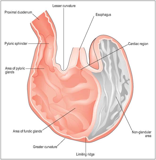
Figure 10.18 • Internal cross-section of rat stomach showing limiting ridge which divides glandular from non-glandular tissue.
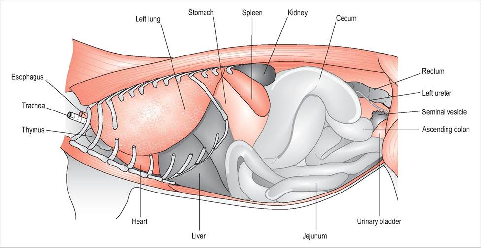
Figure 10.19 • Right lateral thorax and abdomen (some ribs removed). From Popesko, P., Rajtova, V., & Horak, J. (1990) A colour atlas of anatomy of small laboratory animals. Vol. 2. Aylesbury, UK: Wolfe with permission.
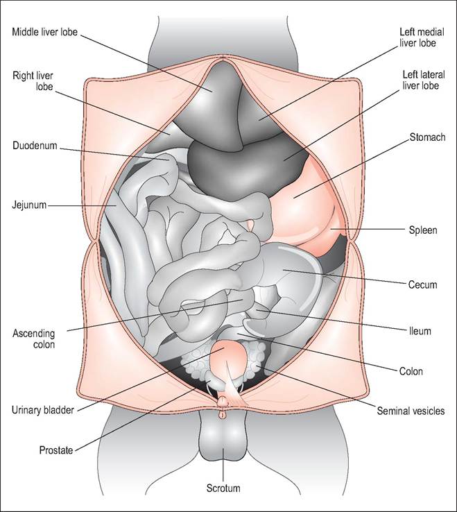
Figure 10.20 • Ventral abdomen of male rat. The cecum of the rat has a very long mesentery so the location can vary. From Popesko, P., Rajtova, V., & Horak, J. (1990) A colour atlas of anatomy of small laboratory animals. Vol. 2. Aylesbury, UK: Wolfe with permission.
darker color and firmer consistency (Hebel & Stromberg 1986c; Sharp & LaRegina 1998).