DIGESTIVE SYSTEM
Feeding patterns
Hamsters are omnivorous and coprophagic (Lipman & Foltz 1996). They feed in 5-minute bursts followed by a 2-hour fast (Bivin et al. 1987). Food intake is about 5-7 g daily and water intake is about 10 ml.
The hamster has evolved certain patterns of behavior consistent with a burrowing and hoarding desert animal. Unlike the greedy rat, they do not increase their food intake following periods of fasting; however, they do hoard away more food in case of further deprivation later (Newcomer et al. 1987).Dentition
The oral aperture can stretch from 12 mm vertically to 17 mm horizontally. The dental formula is 1/1,0/0,0/0,3/3 (Bivin et al. 1987; Lipman & Foltz 1996). The hamster has open-rooted yellow incisors and rooted (brachiodont) molars. The shorter upper incisors can be replaced in 1 week whereas the longer lower incisors take 2.5-3 weeks to regrow. The diastema is longer in the maxilla than the mandible (Figs. 11.4 and 11.5). The mandibular symphysis is freely movable and may not fuse (Harkness & Wagner 1995).
Hamsters (Cricetidae) differ from rats and mice (Muridae) in having molar cusps in two parallel longitudinal rows instead of being arranged in three rows (Derrell Clark 1987). The crowns allow retention of food, which makes hamsters, like humans, susceptible to caries. Male hamsters are more prone to caries than females (Bivin et al. 1987).
The tongue is well developed and very flexible. The muscular bulge at the base contains the small hyoid bone. There are four types of lingual papillae: filiform, fungiform, foliate, and vallate. The major salivary glands are submaxillary, parotid, and sublingual (Bivin et al. 1987; Magalhaes 1968).
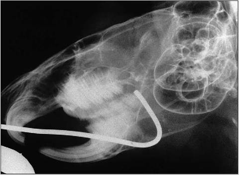
Figure 11.4 • Radiograph of hamster that got the wire of it’s water bottle trapped in the pouch.
Note the large tympanic bulla typical of a nocturnal species.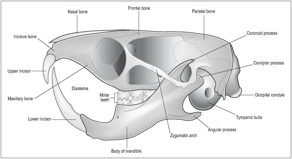
Figure 11.5 • Skull of Syrian hamster (Mesocricetus auratus). From Popesko, P., Rajtova, V., & Horak, J. (1990) A colour atlas of anatomy of small laboratory animals. Vol. 2. Aylesbury, UK: Wolfe with permission.
Cheek pouch
The Arabic term for the hamster in its native Syria is “master of the saddle bags” on account of their using their pouches to transport food, bedding, and even young in times of danger (Harkness & Wagner 1995; Lipman & Foltz 1996; Nowak 1999). The pouches are highly distensible invaginations of the lateral buccal epithelium, which extend from the mouth as far as the dorsocaudal scapulae (Harkness & Wagner 1995) (Fig. 11.6).
The pouch measures 4-8 mm wide when empty and 20 mm when full (Bivin et al. 1987). When filled, the pouches cover the parotid gland, masseter muscles, and lateral neck
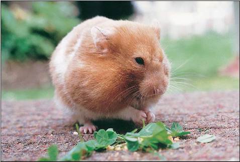
Figure 11.6 • Syrian hamster with bulging cheek pouch.
and shoulder muscles. The mucosa has pale pink folds and is extremely vascular, being supplied by three branches of the external carotid artery. It has, however, no lymphatic supply or adjacent lymph nodes. It is transparent with no hair or glands (Magalhaes 1968).
Cheek pouches are used in research as they are immunologically privileged sites. This is thought to be due to the poor lymphatic blood supply and the lack of glandular tissue. The fact that it can also be easily everted makes it very useful for research on microcirculation, tumors, and transplant surgery (Harkness & Wagner 1995).
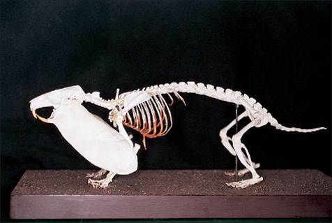
Figure 11.7 • Skeleton of European hamster (Cricetus cricetus) with cast made to show the extent of the cheek pouches. The European hamster is three to four times larger than the Syrian hamster.
Its cheek pouches measure about 60 mm ? 15 mm and have a carrying capacity of 20-30 g!The wall is composed of four layers: keratinized, stratified squamous epithelium, dense collagenous connective tissue, striated muscle fibers, and loose areolar connective tissue where it joins the underlying structures. There are blood vessels and nerves in the connective tissue and muscle. The pouch is emptied by the massaging action of the front feet along with the tongue (Bivin et al. 1987; Magalhaes 1968).
Stomach
The esophagus is lined by keratinized squamous epithelium and leads into the non-glandular part of the stomach. The stomach has two compartments: the non-glandular and the glandular. They are distinguished by a muscular-like sphincter, which may regulate passage of ingesta between the two sections. The esophageal opening lies just cranial to this constriction. The lesser curvature of the stomach is almost non existent as the cardia is located quite near to the pylorus, making vomiting impossible (Hoover et al. 1969; Lipman & Foltz 1996). The total dimensions of the stomach are approximately 3.5?2 cm (Hoover et al. 1969).
Forestomach (non-glandular)
The forestomach is lined by squamous epithelium and a thick muscular layer that is similar to the rumen, although it lacks ruminal papillae. The bacteria are mainly gram-positive, with some gram-negative coliforms (Bivin et al. 1987). The pH is higher than in the glandular stomach, which indicates that mixing of food from both areas does not occur (Hoover et al. 1969).
The presence of volatile fatty acids (mainly acetic) in the forestomach indicates that some fermentation does go on here, with some being absorbed. However, this fermentation role may not be very large as food only stays in the forestomach for about 1 hour (Hoover et al. 1969).
Glandular stomach
This resembles the stomach in the normal monogastric animal and is lined by glandular mucous membranes.
Intestines (Figs. 11.8 and 11.9) defined but often yellow-white in color, owing to associated adipose tissue.