» Digestive System
The esophagus is located on the left side of the neck and is unremarkable, except that it is relatively more deeply placed than in other species and the position of the circular and longitudinal muscle layers are reversed from the typical mammalian pattern; in the camelid there is an inner longitudinal layer and an outer circular layer.
South American camelids have a three-chambered stomach with the chambers identified as C1, C2, and C3. There is no stratification of stomach contents as in ruminants; the ingesta has a consistent composition throughout the chambers and is relatively dry. Figs. 38.21 and 38.22 illustrate the external and internal appearance of the stomach chambers.
All three stomach chambers have glandular saccules, which can develop gastroliths visible on radiographs, and ulcerations can develop in any of the chambers. The most common site for ulceration is at the junction of the nonglandular and true glandular portion of C3. Chambers C1, C2, and the cranial four-fifths of C3 are the sites of anaerobic fermentation of forage. There is a groove that allows milk to bypass the first two chambers when the neonate suckles, although it is not as well defined as the groove found in ruminants.
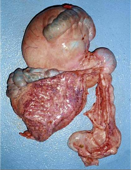
FIG. 38.21 Comparison of the sizes of the large cranial (top left) and caudal (bottom left) sacs of C1 and the smaller, narrower C3 (bottom right). The small C2 makes up the rounded canal in the upper right. The two large dark areas on C1 are the saccular regions. (From Cebra C, Anderson DE, Tibary A, et al: Llama and alpaca care: medicine, surgery, reproduction, nutrition, and herd health, St. Louis, 2014, Elsevier, Fig. 40-5.)
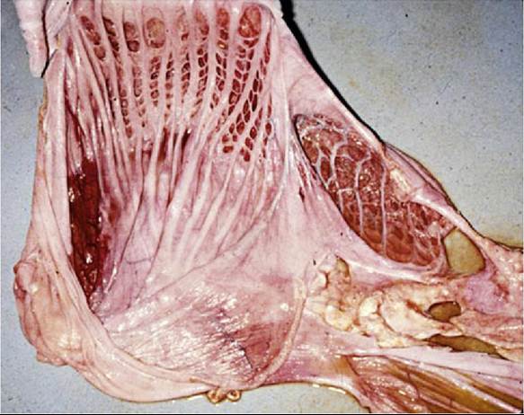
FIG. 38.22 The internal surfaces of C1 (left) and C2 (right).
Note the lack of papillae and the presence of muscular septae dividing the glandular saccules or cells and the wider opening of the second chamber cells. (From Cebra C, Anderson DE, Tibary A, et al: Llama and alpaca care: medicine, surgery, reproduction, nutrition, and herd health,St. Louis, 2014, Elsevier, Fig. 40-6).
The first chamber is by far the largest, holding 83% of the volume, and it is divided into a cranial and caudal sac by a transversely oriented pillar. Most of the surface area of C1 has glandular saccules arranged in rows on the ventral aspect, and only a small portion is covered by stratified squamous epithelium in llamas and alpacas. The glandular saccules contain folds of simple columnar cells that have ultrastructural features indicating both secretory and absorptive function. C1 occupies much of the left side of the abdomen.
Stomach Chamber 1: Distention of C1 causes obvious left-sided abdominal distention. C1 stomach contents are drier than in ruminants, making collection of a fluid sample more challenging. C1 fluid can be obtained through the left flank, midway between the last rib and the stifle. The contractions (three to four per minute; faster if recently fed) cannot be palpated but can be auscultated through the left ventral inguinal area; sounds on the right side are minimal.
The small second chamber holds only 6% of the stomach volume. Its surface contains glandular cells separated by muscular septae, except in the lower curvature where the epithelium is stratified squamous. The mucosa in the glandular cells may be papillated. Despite being referred to as "glandular" saccules and cells, they are not typical stomach glands, and there is little evidence of glandular function of the saccules or cells of C1 and C2, beyond secretion of a thin protective mucous coat. They seem to function by retaining a small volume of stomach contents adjacent to the absorptive cells, with constant turnover from frequent contractions.
The third chamber is long and tubular and contains typical gastric and pyloric glands in its caudal one-fifth.
It is located mainly on the right, and its caudal portion curves upward in the area of the umbilicus. Fig. 38.23 shows the right and left views of the stomach schematically, and Fig. 38.24 illustrates their approximate location within the animal.Passage of a stomach tube should be done via the oral route, because the nasal cavity diameter is usually too small to accommodate a stomach tube. To avoid laceration of the tube by the sharp cheek teeth, a speculum is used. The tube can be palpated in the esophagus on the left side of the neck; if it is not palpable as the tube is advanced, the tube may be in the trachea.
In the camelid neonate the fermentative chambers of the stomach are relatively more developed than those of ruminant neonates. C1 is about 45% of stomach volume at birth and increases to 60% by 6 weeks of age, while the second chamber starts out at about 10% of the stomach volume and gradually declines.
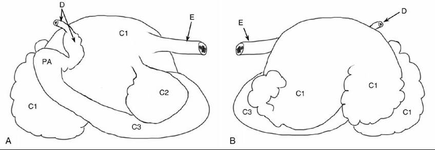
FIG. 38.23 Camelid stomach chamber (schematic). (A) Right side view. (B) Left side view. C1, First chamber; C2, second chamber; C3, third chamber. D, duodenum; E, esophagus; PA, pyloric antrum.
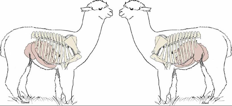
FIG. 38.24 Right and left views of the alpaca illustrating the approximate position of the stomach.
The greater omentum is often translucent because it lacks the extensive fat deposits of the ruminant. It is relatively smaller than that of the ruminant, and there is no omental sling.
The proximal portion of the duodenum has a normal dilatation, referred to as the ampulla, where it connects with and wraps partially around the pylorus in an M-shaped flexure, and then it narrows considerably, making the duodenum one of the common sites for obstruction. The descending duodenum is accessible through the right flank. The mass of the jejunum occupies the right dorsal abdomen.
Most of the jejunum is coiled tightly, with a short mesentery that prevents exteriorization, but the distal one-third has a looser mesentery that allows more mobility. The small intestine in llamas, except for the ampulla of the duodenum, is about 2 cm in diameter in the adult, and in the adult alpaca it is about 1 cm in diameter.The cecum is relatively small, and its base is centrally located in the abdomen with the apex pointing caudally. The spiral colon is similar to that of the ruminant but with more coils and a large, circular proximal loop. The spiral colon is usually caudal to the stomach chambers in the ventral abdomen, but it may be found in the right lateral abdomen. Fig. 38.25 shows the small and large intestine of a llama. Normal feces are formed into pellets that either are separate or are clumped but easily separated.
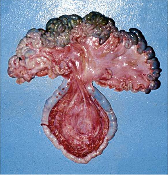
FIG. 38.25 Small intestine (top) and ascending colon (bottom). The mesentery of the ascending colon is long and pendulous; this section of the intestines can most easily become torsed or entrapped. (From Cebra
C, Anderson DE, Tibary A, et al: Llama and alpaca care: medicine, surgery, reproduction, nutrition, and herd health, St. Louis, 2014,
Elsevier, Fig. 40-8.)
In the spiral colon, camelids have an increased chance of obstruction from a foreign body compared to ruminants, because the diameter of the colon decreases from about 5 cm at the beginning of the loops to about 2 cm at the first centripetal coil. Hairballs are not uncommon in young camelids. Because the spiral colon has a relatively loose mesenteric attachment, it can be entirely exteriorized from either a midventral or right paralumbar incision.
The descending colon has a short mesentery; this needs to be noted when performing rectal palpations, which are possible in adult llamas if the examiner has small hands and arms.
The shape and right-sided position of the liver of camelids is very similar to that of ruminants, although camelids usually lack a gallbladder. The liver covers C2 and most of C3, except for a small portion ventrally. Camelids have right, left, caudate, and quadrate lobes of the liver and a fimbriated caudal border.
The preferred site for a liver biopsy is at the right ninth intercostal space, about 20 to 22 cm below the topline.
The pancreas is long and Y-shaped and runs from the area of the common bile duct caudodorsally along the mesentery of the duodenum.