Each Type of Blood Vessel Has Physical Properties Suited to Its Particular Function
In a resting animal, at any one moment, about 25% of the blood volume is in the central circulation and about 75% is in the systemic circulation (Tιιble 18-1). Most of the blood in the systemic circulation is found in the veins.
Only 20% of the systemic blood is found in the arteries, arterioles, and capillaries. Therefore, systemic veins arc known as the blood reservoirs of the circulation. Arteries function as high-pressure conduits for rapid distribution of blood to the various organs. Arterioles are the "gates" of the systemic circulation; they constrict or dilate to control the blood flow to each capillary bed. Although only a small fraction of the systemic blood is found in capillaries at any one time, it is within these exchange vessels that the important diffusional exchange takes place between the blood stream and the interstitial fluid.Table 18-2
Geometry of Systemic Circulation of a 30-kg Resting Dog
| Vessel | Number | Inside diameter (mm) | Total cross- sectional area (cm2) | Length (cm) | Velocity of blood flow (cm∕sec) | Mean blood pressure (mm Hg) |
| Aorta | 1 | 20.0 | 3.1 | 40.0 | 13.0 | 98 |
| Small arteries | 45,000 | 0.14 | 6.9 | 1.5 | 6.0 | 90 |
| Arterioles | 20,000,000 | 0.030 | 140.0 | 0.2 | 0.3 | 60 |
| Capillaries | 1,700,000,000 | 0.008 | 830.0 | 0.05 | 0.05 | 18 |
| Venules | 130,000,000 | 0.020 | 420.0 | 0.1 | 0.1 | 12 |
| Small veins | 73,000 | 0.27 | 42.0 | 1.5 | 1.0 | 6 |
| Venae cavae | 2 | 24.0 | 9.0 | 34.0 | 4.5 | 3 |
Modified from MinorWR: Cardiovascular physiology, NewYork, 1990, Oxford University Press.
Table 18-2 compares the various types of vessels in the systemic circulation of a dog. As the aorta branches into progressively smaller vessels, the diameters of the vessels become smaller, but the number of vessels increases. One aorta supplies blood to 45,000 terminal arteries, each of which gives rise to more than 400 arterioles. Each arteriole typically branches into about 80 capillaries. The capillaries are so small in diameter that red blood cells must pass through in single file. However, because of the sheer number of capillaries, the total cross- sectional area of the capillaries is much greater than the cross- sectional area of the preceding arteries and arterioles. Because capillary blood flow is spread out over such a large cross- sectional area, the flow velocity is low. Blood moves rapidly (about 13 cm∕sec) through the aorta and large arteries. At this speed, blood is delivered from the heart to all parts of the body in less than 10 seconds. The velocity of blood flow decreases as the blood leaves arteries and enters arterioles and capillaries in each tissue. The velocity of blood flow in capillaries is so slow that blood typically takes about 1 second to travel the 0.5-mm length of a capillary. During this time, diffusional exchange takes place between the capillary blood and the interstitial fluid. Blood from the capillaries is collected by venules and veins and is carried quite rapidly back to the heart.
Figure 18-4 depicts the branching pattern in the systemic circulation and graphs the velocity of blood flow within the different types of vessels. This figure emphasizes the rapidity of bulk flow through large vessels and the relatively slow flow through the capillaries. Note that the velocity of blood flow, not the total flow per minute, is lower in the capillaries. The same volume of blood necessarily flows each minute through an artery, the capillaries that it feeds, and the veins draining the capillaries.
In addition to having a large cross-sectional area (and therefore slow velocity of blood flow), capillaries have a large surface area.
The total surface area of the walls of all the capillaries in the systemic circulation of a large dog is about 20 nr, which is nearly 30 times greater than the dog’s body surface area. The large surface area of capillaries helps promote efficient diffusional exchange between the capillary blood and the interstitial fluid.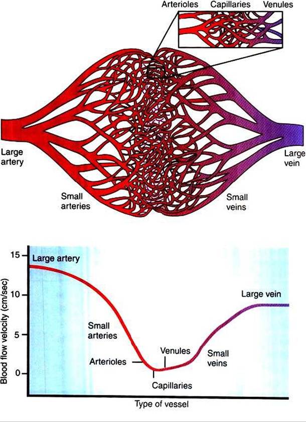
FIGURE 18-4 As the systemic arteries branch to form small arteries, arterioles, and capillaries (toph the total cross-sectional area of the vessels increases, so the forward velocity of blood flow decreases (bottom). As blood from the capillaries is collected into venules and veins, the total cross-sectional area is reduced, so the velocity of blood flow increases again.Therefore, blood moves quickly from the heart to ‘.he microvessels, where it stays for a few seconds before moving rapidly back to the heart.
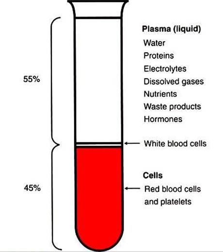
FIGURE 18-5 Anticoagulated blood can be separated into a liquid component (plasma) and a cellular component (cells) by centrifugation. Plasma is a solution of many important substances in water.The presence of proteins gives plasma its typical pale-yellow color.The cells are heavier than the plasma, and they settle to the bottom. Most of the cells are red blood cells. The white blood cells are slightly lighter in weight than the red blood cells, and they form a thin buffy coat on the top of the red cell layer. The fraction of cells in blood is called the hematocrit. In this example the hematocrit is 45%.