Efficient Milk Removal Requires the Release of OxytocinrWhich Causes Contraction of Muscle Cells That Surround the Alveoli (Myoepithelial Cells), and Movement of Milk into the Ducts and Cisterns
To facilitate the process of milk removal, myoeρitheliιιl cells surround the alveoli and ducts (see Figures 39-1 and 39-7). The myoepithelial cells are particularly responsive to oxytocin and, in fact, contract when exposed to the hormone.
The synthesis and release of oxytocin from the posterior pituitary is elicited by a neuroendocrine reflex involving tactile stimulation of the udder by suckling by the young, or the manual stimulation of washing before milking. The sensory stimuli from the udder are carried through the spinal cord into the hypothalamus. Neurons in the paraventricular and supraoptic nuclei are stimulated to synthesize oxytocin and release it from nerve terminals that impinge on the median eminence (Figure 39-8). Other sensory stimuli that elicit oxytocin release include auditory, visual, and olfactory stimuli that occur near or within the kennel, cattery, or milking parlor. Past societies used various deceptions to have earlier breeds of cattle release their milk. Fhey often allowed the calf to suckle one teat while they milked the other glands. They also knew about the Ferguson reflex, if not in name, in which stimulation of the cervix (and release of oxytocin) was elicited by blowing air into the vagina using hollow tubes.The release of oxytocin occurs within seconds after the stimulus arrives in the hypothalamus; increased pressure within the mammary gland is evident within a minute of stimulation as milk is forced out of the alveoli and ducts because of contraction of the myoepithelial cells. The term used in mammals to describe this phenomenon is milk letdown. Increased pressure within the udder is often obvious within a minute of the stimulation. 'Fhe release of oxytocin lasts only a few minutes, and it is important that the milking process begin soon after milk letdown is complete (Figure 39-9). The milking process, as done by machine or by hand in earlier times, is often completed within 4 to 5 minutes.
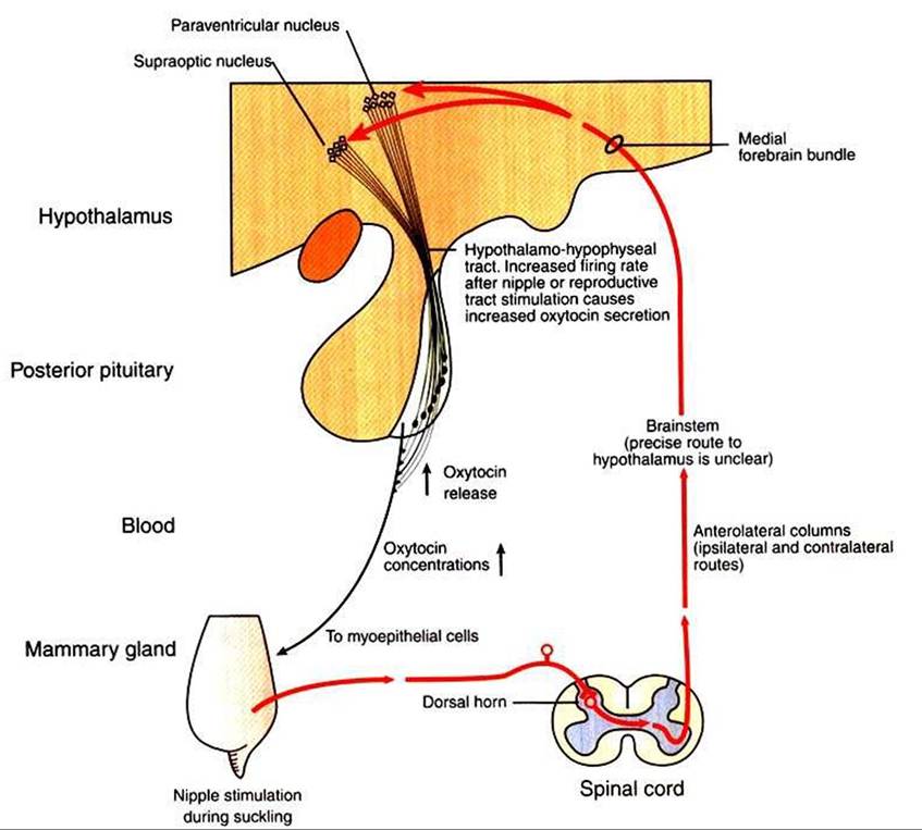
FIGURE 39-8 Somatosensory pathways in the suckling-induced reflex release of oxytocin.
The actual pathway of sensory input in the hypothalamus is unknown, but it probably involves the medial forebrain bundle. (Modified from Johnson M, Everitt B: Essential reproduction, ed 3, London, 1988, Blackwell Scientific.)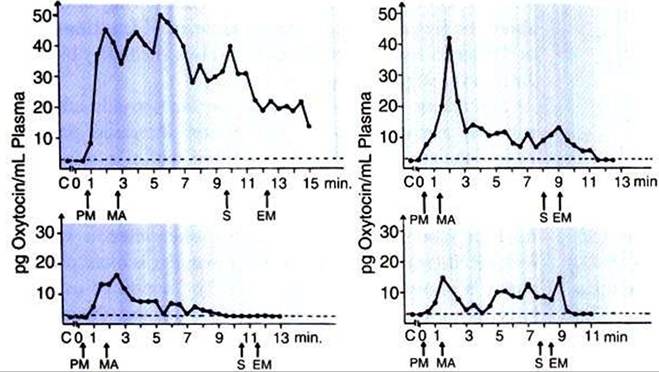
FIGURE 39-9 Oxytocin in the blood of cows before, during, and after milking. Abscissae show time in minutes. C, Control level; EM, end of machine milking; PM, preparation for milking; MA, application of teat cups; S, stripping. (From Cowie AT: Lactation. In Austin CR, Short RV, editors: Reproduction in mammals, ed 2, vol 3, Hormonal control of reproduction, Cambridge, UK, 1984, Cambridge University Press.)
It is interesting to compare stimuli that release oxytocin, which initiates the passive part of lactogenesis, with stimuli that release prolactin, which directly influences lactogenesis. Any sensory stimulus that a cow associates with milking has the potential for releasing oxytocin. The neuroendocrine reflex is elicited in the expectation of milk removal because of the environment (kennel, cattery, or milking parlor) to which the animal is exposed. Prolactin, on the other hand, is released only by tactile stimulation of the udder. The latter makes sense, because there is no need to stimulate milk synthesis and release unless the evidence for milk removal (udder stimulation) is strong. Milk removed during hand milking is trapped in the teat and forced out, whereas milk removed by milking machines moves by suction.