Epaxial Muscles
The epaxial muscles (see Fig. 2.22B) are used for intramuscular injections. Less commonly, they must be separated and detached when access to the vertebral column is necessary. The epaxial muscles comprise three longitudinal systems: iliocostalis, longissimus, and transversospinalis.
The hypaxial muscles consist of the longus colli and longus capitis muscles in the cervical and cranial thoracic regions and the psoas muscle in the lumbar region.The splenius muscle is a strong muscle on the dorsolateral aspect of the neck, extending from the withers to the occiput (see Fig. 2.23A/4). It covers the longissimus capitis muscle, the semispinalis capitis muscle, and parts of the spinalis et semispinalis cervicis et thoracis muscle. It originates from the spinocostotransverse fascia, the spinous processes of the first three thoracic vertebrae, and the nuchal ligament and inserts on the nuchal crest and the mastoid process.
The iliocostalis muscle is relatively thin (see Fig. 2.23B/17) and has only lumbar and thoracic parts. Its bundles span several vertebral segments and, in general, run from caudomedial and dorsal to craniolateral and ventral. The muscle is easily identified over the ribs by the glistening tendons. It arises caudally from the wing of the ilium and also by lumbar fascia from the spinous processes of the lumbar vertebrae. The lumbar portion reduces in size cranially and inserts on the last three to four ribs. The thoracic portion arises lateral to the lumbar part but without any sharp demarcation and extends from the twelfth rib to the transverse process of the last cervical vertebrae.
The iliocostalis is lateral to the longissimus system and is covered by the dorsal serratus and the origins of the latissimus and abdominal oblique muscles. The lumbar part of the iliocostalis muscle of the cat is hardly separate from the longissimus.
The longissimus muscle is much thicker than the preceding muscle (see Fig. 2.23B). Its bundles are similarly oriented but are largely fused, giving a uniform appearance to the lumbar and thoracic regions. The thoracolumbar part (longissimus dorsi) is credited with the powerful extension of the vertebral column during the propulsive phase of the gallop. It is related medially to the multifidus, and over the thoracic vertebrae, it is covered dorsally by the spinalis et semispinalis (Fig. 12.10/1 and 2), although it is separated from both by a fibrous septum that serves as the origin of the last- named muscle. The ventral edge of this septum ends near the transverse processes of the vertebrae and is a landmark in the surgical approach to the intervertebral disks.
The lumbar part of the longissimus muscle arises from the wing of the ilium and the lumbar spinous processes, against which it lies. Along its length it detaches several bundles, arranged in a lateral and a medial row, which cover the bases of the lumbar transverse processes before ending on accessory processes of the cranial six lumbar vertebrae. The caudal narrow part, not covered by the middle gluteus, inserts dorsally mainly on the arch of the last lumbar vertebra and the last intervertebral disk, with more limited insertion on the sixth and fifth lumbar vertebrae. The longissimus lumborum is covered by a dense aponeurosis separated from the thoracolumbar fascia by fat.
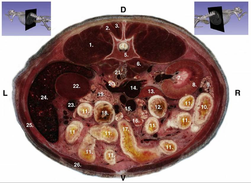
FIG. 12.11 Transverse section of the dog at the level of the 2nd lumbar vertebra. 1, Iliocostalis and longissimus muscles; 2, Spinalis and semispinalis muscle; 3, Multifidi muscles; 4, Spinal cord; 5, 2nd lumbar vertebra; 6, Psoas muscles; 7, Renal medulla (renal papilla); 8, Renal cortex; 9, Pancreas, right lobe; 10, Descending duodenum; 11, Jejunum; 12, Ascending colon; 13, Liver (caudate process of the caudal lobe); 14, Caudal vena cava; 15, Cranial mesenteric artery and vein; 16, Mesenteric lymph nodes;
17, Duodenojejunal flexure (terminal part of the ascending duodenum); 18, Descending colon; 19, Pancreas, left lobe; 20, Left adrenal gland; 21, Abdominal aorta; 22, Left kidney (cranial pole); 23, Vessels of the spleen; 24, Spleen; 25, Internal, external obliquus and transversus abdominalis muscles; 26, Rectus abdominis muscle.
The thoracic part (see Fig. 2.23B/16") inserts by medial tendons on the transverse or accessory processes of the thoracic vertebrae and by lateral tendons on the necks of the last seven ribs. The dorsal branches of the thoracic nerves pass between the medial and lateral tendon.
The cervical part (see Fig. 2.23B/16') of the longissimus muscle has a triangular form, filling up the angle between the cervical and thoracic vertebrae, and comprises four incompletely separable bundles, which arise from the transverse and articular processes of the first thoracic vertebrae and insert on the transverse processes of the sixth to third cervical vertebrae.
The longissimus capitis muscle, strong and flat, lies medial to the longissimus cervicis and splenius muscles (see Fig. 2.23/16'). It originates from the transverse processes of the first three thoracic vertebrae and from the caudal articular processes of the last three or four cervical vertebrae. It runs over the dorsal surface of the atlas and inserts on the mastoid process, fused at the level of the atlas with the splenius muscle.
The longissimus atlantis muscle, present in only 20% of dogs, arises from the articular processes of the last three cervical vertebrae and ends on the wing of the atlas.
In the cat there is a longissimus capitis but not a longissimus atlantis. Furthermore, it is not possible to separate the cervical and thoracic longissimus muscles; a shallow longitudinal groove appears to separate the lumbar portion into lateral and medial parts.
The more complex transversospinalis system is more intimately related to the vertebrae. Some fascicles connect one vertebra to the next, whereas others span several vertebrae; most are oriented from caudoventral and lateral to craniodorsal and medial, in contrast to the direction taken by the preceding muscles. The transversospinalis system comprises the spinalis et semispinalis thoracis et cervicis, semispinalis capitis, and several less important, more obviously segmental muscles (multifidi, intertransversarii, interspinales, and rotatores) that lie directly on the vertebrae (see Fig.
2.23B/15).The spinalis et semispinalis thoracis et cervicis muscles extend from the midlumbar region to the spine of the axis and lie against the lateral surface of the spinous processes (see Fig. 2.24A/2 " and 2'' ') dorsomedial to the longissimus thoracis. Their fascicles connect spinous and mammillary processes with more cranial spinous processes. They are a powerful muscle incompletely divided into a lateral part, the spinalis et semispinalis thoracis, and a medial part, the spinalis cervicis.
The spinalis et semispinalis thoracis (lateral part) arises from the aponeurosis of the thoracic part of the longissimus muscle and from the midlumbar spinous processes. It inserts on the spinous processes of the sixth thoracic forward to the sixth cervical vertebrae. The tendons that are attached to the last two cervical spines are particularly powerful and form a plate that is fused with the tendon of the medial part of the muscle. In the cat, the spinalis et semispinalis thoracis arises from only the tenth or eleventh thoracic spinous process forward.
The spinalis cervicis (medial part) lies dorsomedial to the lateral part. It arises from the spinous processes of the sixth to first thoracic vertebrae and continues as a flat muscle with four tendinous inscriptions in its belly on the spinous processes of the fifth to second cervical vertebrae.
The semispinalis capitis is a more independent neck muscle lying between the splenius and the cervical components of the preceding muscle (see Fig. 2.23B/15). It is clearly divided into the biventer cervicis and the complexus, which both contact their fellows and the ligamentum nuchae in the median plane. The biventer is the more dorsal and more caudal of the two. It arises from and around the transverse processes of the first few thoracic vertebrae, medial to the longissimus cervicis and capitis, and ends on the occipital bone ventral to the external occipital protuberance. It can be identified by several tendinous inscriptions.
The complexus arises from the articular processes of the caudal four cervical vertebrae and first thoracic vertebra and ends on the nuchal crest; it is not segmented.In the cat, the biventer is relatively poorly developed, and it has only two to three tendinous inscriptions. The complexus, even less developed, is divided by a distinct strip of tendon that runs horizontally through its middle.
The multifidus is more distinctly segmented in the dog than in the cat, especially in the cervical region. The lumbar part in the dog consists of 10 to 11 bundles that arise from the mammillary process of the first caudal vertebra, the rudimentary articular processes of the sacrum, and the mammillary processes of the lumbar vertebrae and last two thoracic vertebrae (see Fig. 2.24B/2'). As a rule, two segments are passed by each bundle: thus the insertions are to the spinous processes of the sixth lumbar to the tenth thoracic vertebrae. The thoracic part comprises nine distinct bundles that arise from the mammillary and transverse processes of most thoracic vertebrae and, after passing two segments, insert on the spinous processes of the first eight thoracic and last cervical vertebrae.
The cervical part, which is completely covered by the semispinalis capitis, consists of six individual parts that divide into large lateral and smaller medial bundles.
The intertransversarii muscles, divisible into lumbar, thoracic, and cervical units, are sometimes regarded as being split from the longissimus system. The lumbar units are especially well developed in cats, whereas in dogs the muscle is composed of thin bundles. In both species they unite the mammillary and accessory processes of the lumbar vertebrae and last four thoracic vertebrae with the transverse processes of the twelfth to sixth thoracic vertebrae, never passing more than three segments.
The cervical intertransversarii are much stronger and are arranged in dorsal, middle, and ventral layers. The dorsal layer is located between the insertions of the longissimus cervicis and capitis muscles and comprises five bundles only partly separable.
The middle layer consists of five to six thin, separate parts, of which the deeper fibers run from segment to segment and the superficial ones always pass over one segment. The ventral layer lies dorsal to the longus capitis. It arises from the ventral border, the transverse process of the sixth cervical vertebra, and runs forward, as three digitations, to processes of the fourth to second vertebrae.The interspinales muscles connect the spinous processes of the vertebrae in the lumbar, thoracic, and cervical regions; the lumbar portions are completely covered by the multifidus. The thoracic parts are broader.
Deep rotator muscles lie medial to the multifidi in the cranial thoracic region. The eight long rotators extend between the transverse and spinous, spanning two joints; the nine short ones pass between adjacent vertebrae. It is not possible to separate the rotator muscles from the multifidus in the cat.