EXTERNAL FEATURES AND INTEGUMENT
Feathers provide the principal characteristic that distinguishes birds from mammals. They streamline the body and assist in transforming the forelimbs into wings. The
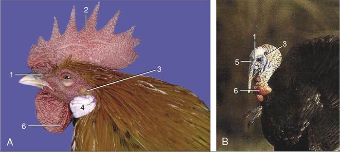
Figure 37-1 Head of the chicken (A) and the turkey (B).
1, Nostril; 2, comb; 3, ear opening; 4, ear lobes; 5, snood; 6, wattles.
Figure 37-2 A, B, Two chickens with ornaments. 1, Comb; 2, wattles; 3, ear lobe.
feathers are among the features (others are mentioned later) that lighten birds relative to their size and thus enhance their efficiency in the air. Feathers have many functions that in mammals are performed by hairy skin: protection against mechanical, radiological, thermal, chemical, and biological influences, and thermoregulation, and communication.
The skin is thin, loose, and tears easily; however, because it is poorly supplied with blood vessels and nerves, wounds do not bleed as much as in mammals, and birds seem relatively insensitive to manipulation of their skin. The skin in chickens is yellowish over the body but may be more deeply pigmented on the shanks and feet. It is paler in productive laying hens, in which the pigment is withdrawn and incorporated in the yolk. The dorsal surface of the neck-trunk junction is recommended for subcutaneous injections. Other locations are the cranial skin fold of the knee and the lateral side of the thorax. In most species, including the domestic chicken, localized changes in the skin occur during the brooding period for the more efficient incubation of the eggs. Brooding (incubation) patches that develop on the breast are characterized by feather loss and by thickening, edema, and increased vascularity.
The subcutaneous layer is mainly composed of loose connective tissue; it also contains fat, most copiously present in aquatic and arctic species like penguins, ducks, geese, and swans, and in migratory species before migration.
The comb, wattles, and ear lobes (and the snood of turkeys) are soft ornamental outgrowths of the skin about the head (Figure 37-1, A-B, and Figure 37-2, A-B). Their dermis is thick and vascular, but the covering epidermis is thin. They are thus easily injured and provide potential portals for infection. In nearly all commercially reared chicks the comb (and snood) are snipped off (dubbing, desnooding) to prevent their traumatization in the confined spaces in which these birds are held. The edges of the wattles are used for intradermal injections.
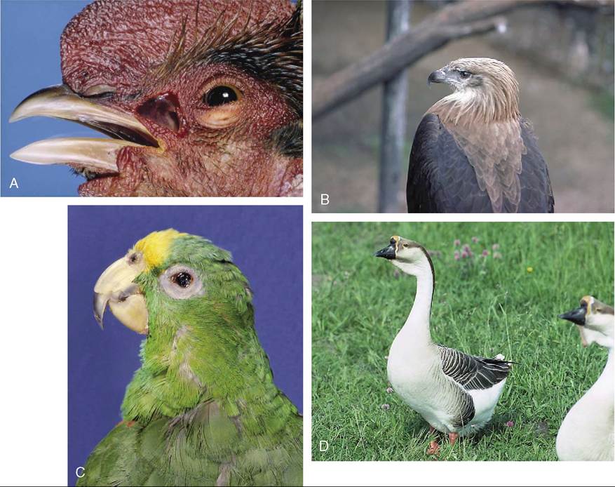
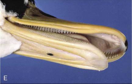
Figure 37-3 Differences in the form of the avian head (A-D). In E, the filter mechanism in the beak of a duck.
The beak (bill) is the functional counterpart of the lips and teeth of mammals. It is a derivative of the skin and provides a horny cover (rhamphotheca) for the rostral parts of both upper (rhinotheca) and lower (gnathotheca) jaws that grows continuously to compensate for natural wear. The beak varies tremendously in form among species, according to diet (Figure 37-3, A-E). A rich innervation causes it to be quite sensitive. Most commercially raised chickens and turkeys are debeaked when young (cutting off the upper beak in front of the nostrils) to prevent cannibalism. In psitta- cines, pigeons, and raptors, the base of the maxillary rhamphotheca, called the cere, may enclose the nostrils (Figure 37-3, C-D).
It is composed of softer keratin than the rest and is particularly prominent and fleshy in waterfowl as well as in budgerigars, in which it is used as a guide to their sex; the cere of the cock is blue, and that of the hen is light brownish pink.
The scales on the shanks and feet are cornified epidermal patches similar to those of reptiles (Figure 37-4, A-B). The feet of most birds are adapted for perching or holding prey and have one toe facing backwards, three facing forwards (anisodactyl).
In waterfowl the three forward-pointing toes are connected by skin (webbed) to make more efficient sculls (palmate). Some species, like psittacines, have two (first and fourth) toes facing backward and two (second and third) facing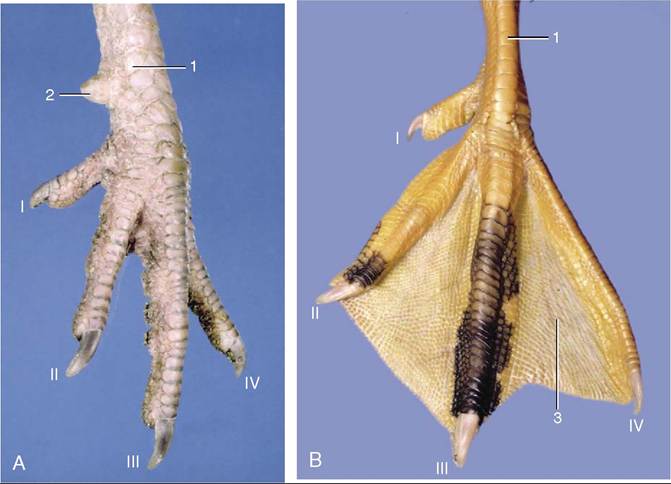
Figure 37-4 Left foot of a cockerel (A). Left foot of a goose (B). 1, Shank (metatarsus); 2, spur; 3, web between toes; I-V toes.
forward (zygodactyl): these species use their feet for grasping and climbing. The spur developed on the cau- domedial surface of the rooster’s shank is used as a weapon; it has an osseous core within a cone of horn. The length of the spur and the growth rings at its base may be used for determining age. Removal of the spur papilla in the chick inhibits its growth, much as the removal of the horn bud prevents horn growth in ruminants.
There are only three discrete skin glands: the sebaceous uropygial gland (preen or oil gland; Figure 37-5), the aural gland, and the vent gland. The absence of sweat glands means that the birds have to lose heat through their skin and by evaporation from the respiratory system. The epidermis has the unique feature that allows it to act like a holocrine sebaceous gland, secreting a thin lipid film that helps in the maintenances of the plumage.
The uropygial gland in chickens is bilobed, about 2 cm in diameter, and located dorsal to the vertebrae that form the short tail. Its fatty secretion emerges from paired openings atop a small cutaneous papilla. The lipid secretion is carried to the body and wing feathers during preening. In waterfowl the secretion is important for waterproofing the feathers and insulating the submerged part of the body. This lipid layer also forms a protective bacteriostatic layer that may explain why birds are little prone to skin infections. The uropygial gland is prominent in budgerigars and African greys but absent from many other parrots (e.g., Amazon parrots), ostriches, and many pigeons.
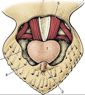
Figure 37-5 Uropygial (preen) gland; dorsal view. 1, Uro- pygial gland; 2, papilla of uropygial gland through which the secretion is extruded; 3, cut edge of skin; 4, feather follicle; 5, caudal vertebrae and associated muscles.
Aural sebaceous glands around the external ear secrete a waxy substance. Vent glands secrete mucus; their function is uncertain but may be linked to internal fertilization.