Female Reproductive System
The production of eggs for human consumption is a primary reason for keeping poultry worldwide. Among female birds, both right and left ovaries and oviducts are present embryologically, but in chickens, turkeys, and domestic geese the right organs regress early in development and only the left side develops (both sides persist and develop in ducks).
The ovaries are located cranioventral to the corresponding kidney, and the size varies with the reproductive status of the bird. Prior to the onset of the first laying period, the small ovary is smooth; as puberty is approached, it begins to develop a granular, then cobblestone appearance as follicles enlarge in preparation for ovulation. Just prior to “coming into lay,” the ovary will resemble a bunch of grapes.The mature ovum is more-or-less equivalent to the yolk of the egg; it will be released at ovulation and received into the expanded terminus of the oviduct, the infundibulum (Figure 30-8). It is in the infundibulum that fertilization will take place if the bird is allowed to breed. The oviduct is variable in size, being maximally expanded and lengthened during the lay (60-70 cm long in the hen) and regressing in size during broodiness and the molt. it possesses grossly identifiable sections (the magnum, isthmus, uterus, and vagina) that correlate with the important tasks of producing the albumen (the “white” of the egg) and the laying down of the egg shell. The oviduct terminates in a slitlike aperture in the urodeum, next to the opening of the ureter (Figure 30-8).
Egg Formation and Oviposition
The number of eggs laid consecutively by wild birds during any period of egg laying is termed a clutch. The number of eggs per clutch for a wild bird is probably related to the ability of the species to incubate the eggs and raise the hatchlings. The time period between clutches for wild birds may extend to months or years.
Under typical management practices for flocks of domestic chickens used for egg production, eggs do not remain with hens after the eggs are laid. However, hens typically lay a number of eggs consecutively for several days and then interrupt laying for at least 1 day. some refer to this pattern as a sequence of eggs, rather than a clutch. During a 12-month laying period, hens may produce more than 300 eggs by laying multiple sequences (clutches).The time period between ovulation and ovi- position (expulsion of the egg) for the domestic hen is about 24-26 hours. Ovulation of the next egg occurs after oviposition of the preceding egg, so only one egg is in the oviduct at a time. As in mammals, ovulation is preceded by a sudden, rapid rise in luteinizing hormone (LH), and this hormone surge is believed to be responsible for changes in the mature follicle about to be released from the ovary. Four to 6 hours prior to the LH surge, plasma progesterone levels peak at the highest levels seen in a laying cycle, and this peak appears to be neces-
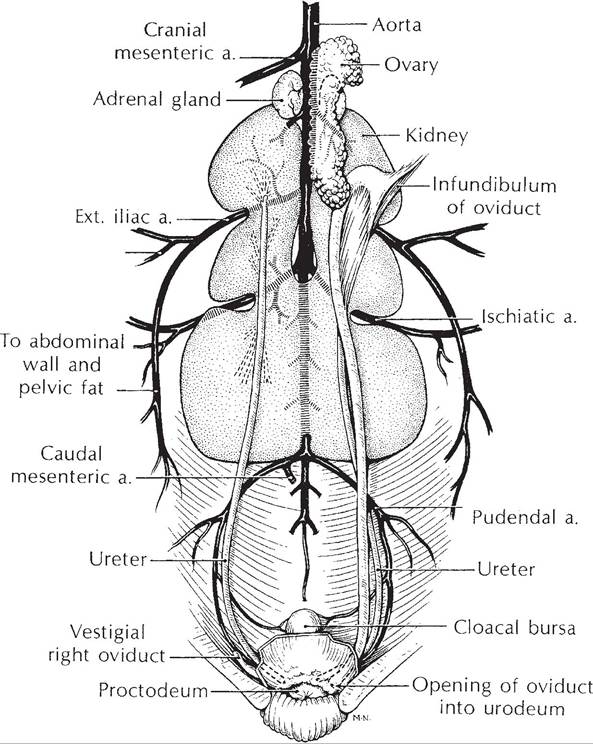
Figure 30-8. The avian genitourinary tract (female). (Reprinted with permission of Wolters Kluwer from Rosskopf,
W. and Woerpel, R. Diseases of Cage and Aviary Birds, 3rd edition. Philadelphia: Williams & Wilkins, 1996.)
sary for the subsequent surge. The source of this progesterone appears to be the largest and most mature ovarian follicle. Unlike mammals, there is no corpus luteum formed on an ovary at the site of an ovulated follicle. Progesterone receptors are found in various sites throughout the oviduct, where progesterone may promote secretions and muscular contractions for egg development and transport, respectively.
For fertilization to occur, spermatozoa must reach the egg before it enters the tubular segment of the infundibulum where the first albumen layer will be secreted to surround the egg. Spermatozoa may be temporarily stored in the infundibulum so that they can be readily available for the relatively short period that fertilization is possible.
In the domestic hen, the egg is in the infundibulum for about 15 minutes. Spermatozoa are also stored in specialized tubules in the uterovaginal region for longer periods. In the domestic hen, spermatozoa remain viable for 7-14 days when stored here. Spermatozoa are released from this site in association with ovipostion of each egg and migrate to the infundibulum to be available to fertilize the next egg. Ovulation of the next egg occurs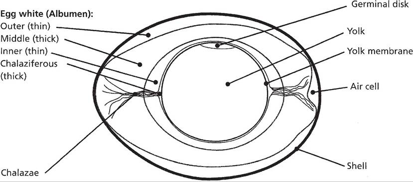
Figure 30-9. Basic internal structure of a typical egg. (Reprinted with permission of Wiley-Blackwell from Reece, W.O. Functional Anatomy and Physiology of Domestic Animals, 3rd ed. Baltimore: Lippincott, Williams & Wilkins, 2005.)
about 30-45 minutes after oviposition of the previous egg.
Albumen (egg white) secretion around the yolk begins in the infundibulum, but the majority is secreted by the magnum. Albumen primarily consists of water, protein, and minerals and is a source of these nutrients for the developing embryo. some of the proteins also have antimicrobial properties and protect against microorganisms. The chalazae are two twisted, fiberlike structures that extend from opposite sides of the yolk to each end of the egg (Fig. 30-9). These are formed from fibers in the inner layers of the albumen when the yolk rotates as it passes through the oviduct.
The eggshell consists of four layers that were applied to the outer surface of the albumen. From inner to outer, the layers are (1) shell membranes, (2) mammillary cores, (3) matrix, and (4) cuticle. The combination of the mammillary cores and matrix has also been termed the testa. The innermost shell membranes permit the exchange of gases and water, but these membranes are not permeable to albumen. As eggs age, the inner and outer shell membranes tend to separate at the large end of the egg, and an air cell is formed (Fig. 30-9). The size of an air cell increases as an egg continues to age.
Egg candling, shining a beam of light through the intact egg to view the interior, is used to determine the size of the air cell, and this is one measure of egg quality.The mineral component of the eggshell, primarily calcium carbonate, is deposited within the mammillary core and matrix layers. The mammillary cores and matrix layers contain proteins, carbohydrates, and mucopolysaccharides, which provide a framework for the deposition of calcium carbonate crystals (i.e., calcification). several different proteins that contribute to this framework have been identified, and experiments suggest that the appropriate production of these proteins by the shell gland is a key factor in determining shell quality. Gene expression studies indicate that the production of these proteins is influenced by hormonal factors, such as progesterone, and that rates of gene expression vary greatly as the egg passes through the shell gland. The outermost cuticle layer contains lipid, prevents evaporation, is water repellent, and functions as a barrier to microorganisms.
Eggshell formation in the uterus or shell gland of the domestic chicken requires about 17-20 hours. During this period, approximately 2-2.5 grams of calcium are transported from the blood by the uterus and deposited in the eggshell as calcium carbonate crystals (calcite). Approximately 95% of the total weight of an eggshell is calcium carbonate. Plasma calcium levels decrease during the period of eggshell formation and then rebound before the next period of eggshell formation begins. During periods of active eggshell formation, calcium is added to the blood by intestinal absorption and mobilization of medullary bone. If dietary calcium is adequate, the majority of the necessary calcium can be obtained during this period by intestinal absorption. However, a net loss of medullary bone calcium will occur during the hours of active eggshell formation, even with adequate calcium intakes. Replacement of the lost medullary bone can be done with calcium gained by intestinal absorption during the hours that active eggshell formation is not occurring.
Vitamin D stimulates both the intestinal absorption of calcium and transport of calcium by the shell gland (uterus). Parathyroid hormone also promotes calcium transport by the shell gland for eggshell formation.Osteoclasts are responsible for the mobilization of calcium from medullary bone, so the increased mobilization during eggshell formation reflects an increased osteoclastic activity. Osteoclasts also mobilize calcium from lamellar cortical bone throughout the skeleton, and a net loss of calcium from cortical bone does occur in high-producing laying chickens who have been laying for extended periods. Osteoporosis, a progressive decrease in mineralized bone leading to bone fragility and increased risk for fractures, is recognized in laying flocks and is believed to contribute to the syndrome cage layer fatigue, in which birds are found paralyzed in their cage. Numerous fractures may be found involving the ribs and long bones of such birds.
Calcium carbonate deposition within the eggshell also requires the localized formation of carbonate ions within the shell gland. This generation of carbonate ions is dependent on carbonic anhydrase activity within the gland. Under the influence of carbonic anhydrase, CO2 from blood perfusing the gland is used to produce carbonate ions. Laying chickens subjected to abnormally high environmental temperatures (heat stress) typically have declines in egg production and in the shell quality of eggs that are produced. The decline in shell quality is associated with a reduction in blood PCO2, as a result of hyperventilation induced by heat stress. The lower CO2 is believed to adversely affect the ability of the shell gland to produce carbonate ions. Heat-stressed birds also reduce food intake, which would also contribute to the decline in egg quantity and quality.
Arginine vasotocin (AVT) stimulates uterine contractions during oviposition. Like oxytocin in mammals, AVT is released from the posterior pituitary, and its plasma levels rise significantly shortly before and peak during oviposition. Unlike mammals, birds have one small peptide hormone, AVT, that functions in both the regulation of urine osmolality and the regulation of uterine contractions.