Fertilization
Spermatozoa Transport and Viability
In most cases, fertilization occurs in the uterine tube next to the ovary; during natural copulation, spermatozoa are deposited in the vagina (most species) or uterus via the cervix (mare,
sow, and bitch).
Even though ejaculated spermatozoa are motile, the major factor in the transport of spermatozoa to the site of fertilization is muscular activity of the tubular genitalia following insemination. The time required for spermatozoa to travel to the site of fertilization in a cow is about 2.5 minutes, but the first arriving spermatozoa typically do not accomplish fertilization. Based on calculated speed of bull spermatozoa, it would take 1.5 hours for them to swim this distance. Less than 0.5% of the ejaculated spermatozoa reach the site of fertilization.Oxytocin, a peptide hormone from the neurohypophysis (see Chapter 12), promotes muscular activity of the female tubular genitalia to assist with spermatozoa transport. it is released in the cow during natural mating and during artificial insemination, presumably as a result of a neural reflex initiated by physical stimulation of the female tract.
spermatozoa must remain in the female reproductive tract for some period after ejaculation before they are capable of fertilization. The process that occurs here to convert nonfertile spermatozoa to fertile spermatozoa is termed capacitation. Capacitation includes changes in or removal of components of the outer acrosome and plasma membranes so that acrosomal enzymes can later be released and activated. Part of the natural capacitation process requires exposure of the spermatozoa to female reproductive tract secretions, but capacitation of spermatozoa can be done in vitro using experimentally derived protocols and solutions.
Under normal conditions, viability and survival times of spermatozoa in the female reproductive tract are only a matter of hours.
Length of fertility in the female tract is as follows: ewe, 30 to 48 hours; cow, 28 to 50 hours; mare, 144 hours. Motility may last somewhat longer than fertility. The limited viability of spermatozoa means that insemination must occur within hours of ovulation so that viable spermatozoa are present when ova arrive for fertilization. in most species, female sexual receptivity begins some hours prior to ovulation, so that this is possible.Gamete Fusion and Early Embryonic Development
At ovulation, a zona pellucida, a relatively thick membranous structure consisting of crosslinked glycoproteins, surrounds the vitelline membrane (cell membrane or plasma membrane) of the ovum. in most cases, a variable number of granulosa cells surround the zona, and this layer is termed the cumulus oopho- rous. The zona is believed to be a product of the innermost layer of granulosa cells. Microvilli from the vitelline membrane of the ovum penetrate the zona, as do processes from the granulosa cells. The first polar body, which results from the first meiotic division, also accompanies the ovulated ovum within the zona (see Chapter 27).
The zona pellucida is a semipermeable membrane that helps protect the ovum and that has receptor sites for attachment of spermatozoa during fertilization. A specific protein, ZP3, in the zona serves as a binding site for spermatozoa. The precise structure of this protein varies among species, and this variation is the primary reason that spermatozoa from one species cannot bind to and fertilize ova from other species.
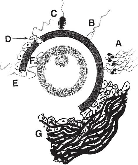
During or just after binding and attachment to the zona, spermatozoa undergo a series of events termed the acrosome reaction (Fig. 281). As part of this reaction, locally released acrosome enzymes digest a passage through the zona. This passage permits spermatozoa to swim their way to the vitelline membrane of the ovum, which is accomplished in a matter of minutes. Multiple spermatozoa may attach to the zona of a single ovum, even though only one spermatozoon will ultimately be responsible for fertilization.
After penetration of the zona, the cell membrane of the single spermatozoon that will accomplish fertilization attaches to and fuses with the vitelline membrane of the ovum (Fig. 28-1). This initiates the second meiotic division by the ovum, which results in formation of the second polar body. The fusion of the spermatozoon with the ovum also stimulates release of cytoplasmic granules by the ovum, whose con-
Figure 28-1. Sequence of events from initial binding of spermatozoon to zona pellucida to fusion between plasma membranes of spermatozoon and oocyte: A, spermatozoa arrive at site of fertilization; B, initial binding of spermatozoon to zona pellucida; c, acrosome reaction; D and E, penetration of zona pellucida; F, fusion of plasma membranes of spermatozoon and oocytes; G, the cumulus matrix. (Reprinted with permission of Wiley-Blackwell from Hafez E.S.E. and Hafez B. Reproduction in Farm Animals. 7th ed. Philadelphia: Lippincott Williams & Wilkins, 2000.)
tents bring about changes in the chemical nature of the zona pellucida. These changes act to prevent penetration by other spermatozoa. This, together with changes in the vitelline membrane, prevents polyspermy, entry of more than one spermatozoon into the ovum. When these mechanisms fail and polyspermic fertilization occurs, the typical result is early embryonic death. This is believed to be rare in most domestic species when fertilization occurs in vivo, but the incidence of polyspermy is higher when fertilization is done in vitro.
A maternal pronucleus is formed in the ovum by the enclosure of the maternal chromosomes in a nuclear membrane. The head of the sperm enlarges, becoming the male pronucleus. The two pronuclei come together and fuse membranes, forming one cell with the genetic material of both parents. The new cell is ready for cleavage and formation of a morula. cleavage and the subsequent development into a morula and blastula are described in detail in Chapter 3.
During early development, the embryo is not attached to the epithelium lining the female reproductive tract. During this period, embryos obtain their nourishment from fluids and nutrients secreted by glands in the walls of the reproductive organs (e.g., endometrial glands in the walls of the uterus). Progesterone stimulates secretion by these glands, and blood levels of progesterone are relatively high during this period, as it is being secreted from ovarian corpora lutea.
Maternal recognition of pregnancy is detection of a developing embryo, which prevents regression of the progesterone-secreting corpora lutea. Several mechanisms have been identified in different species, but in general, the mechanisms involve secretory products from the developing embryo. These products (e.g., proteins or steroids) act locally within the reproductive tract. in most cases, the embryonic secretory products inhibit the uterine secretion of prostaglandin F2α (PGF2α). Recall that uterine secretion of PGF2α is the key hormonal signal that causes Ieutolysis in most domestic species. In litter-bearing animals, a minimal number of developing embryos appears to be required to recognize pregnancy and prevent regression of the corpora lutea. This number is about four for the sow.
Embryos develop to the blastula stage while still enclosed in a zona pellucida (see Chapter 3). The zona is shed (hatching) prior to attachment of the embryo to the uterine wall for placentation. The outermost layer of cells of the blastula is the trophoblast, and it is from these cells that the fetal membranes will be formed.
Early embryonic death (death of the embryo prior to attachment to the uterine wall) is responsible for a significant number of reproductive failures in domestic animals. Some studies report that up to 30% of fertilized embryos die before developing into fetuses. Possible causes of embryonic death include inherited lethal factors, infections, nutritional deficiencies, inappropriate levels of maternal hormones, and defects in the ovum or spermatozoa before fertilization.