FUNCTIONAL ANATOMY OF THE FEMALE REPRODUCTIVE SYSTEM
1. When conducting a rectal palpation on a cow for the components of the female reproductive system, would one search dorsally (above) or ventrally (below)? What is the relative location of the urinary bladder?
2.
Do all domestic animals (intact females) ovulate over the entire surface of the ovary?3. Compare numbers of spermatozoa and oocytes that develop from one primary spermatocyte and one primary oocyte, respectively.
4. What is the process of oocyte formation known as?
5. What are primordial follicles? Does their number at birth, aside from those destined to become mature oocytes, continue throughout the reproductive life of the female?
6. What function is served by the uterine tubes?
7. What are fimbria?
8. What is the serous covering of the uterine tubes known as?
9. What function is served by the uterus?
0. Is the endometrium glandular throughout in all domestic animals (intact females)?
11. What function is served by the glandular secretion of the endometrium?
2. Is the cervix open at all times?
3. What composes the myometrium and what is its function?
4. What is the major support for the gravid uterus?
L5. What is the landmark junction between the vagina and the vulva? What is the vestibule of the vagina?
6. What is the fornix?
7. What is the major blood supply to the uterus? What is fremitus?
8. What function is served by the intertwining of the uterine artery and vein?
The reproductive system of female domestic mammals consists of two ovaries and the tubular genital tract composed of two uterine tubes, uterus, vagina, and the external genitalia (Figure 15-1). The mammary glands are an important part of the reproductive system as well, and are described separately. The location of the reproductive system relative to the rectum and bladder is shown in Figure 15-2.
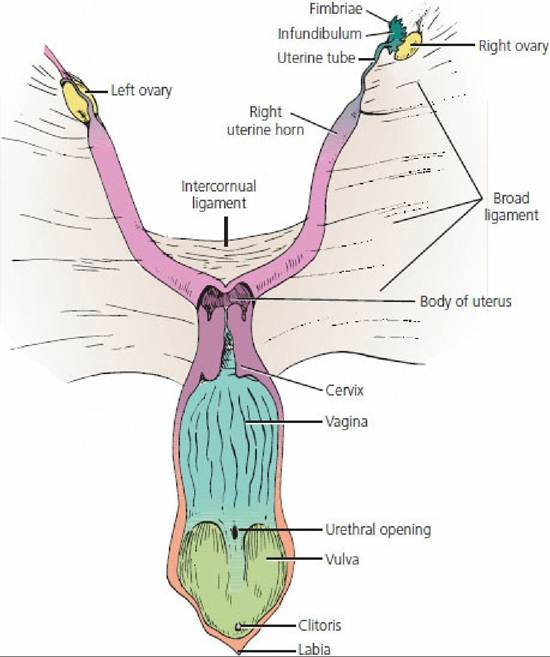
■ FIGURE 15-1 Reproductive tract of the cow (dorsal aspect).
The body of the uterus, vagina, and vulva (vestibule of the vagina) have been laid open and the right ovary withdrawn from the ovarian bursa and infundibulum. The broad ligament (a downward reflection of the peritoneum) suspends the reproductive tract from the dorsolateral abdominal wall.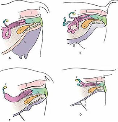
■ FIGURE 15-2 Location of reproductive organs relative to the rectum and urinary bladder. A. Cow. B. Sow. C. Mare. D. Bitch. Note species differences in anatomy of the cervix and mammary gland(s). 1, rectum; 2, urinary bladder; 3, cervix; 4, uterus; 5, vagina; 6, vulva; 7, ovary; 8, mammary gland(s).
Ovaries
The ovaries are paired glands that provide for the development of oocytes and for the production of hormones. Each ovary is located caudal to its respective right or left kidney and is suspended from the dorsal wall of the abdomen by a reflection of the peritoneum, the mesovarium. The mesovarium is part of the broad ligament (Figure 15-3), an inclusive term that also refers to the suspensions of the uterine tubes (mesosalpinx) and uterus (mesometrium). The rather pendulous suspension of the ovaries provides for easy manipulation by rectal palpation in the cow and horse. The ovaries are described as almond shaped in most species and as bean shaped (kidney shaped) in the mare (Figure 15-4). In the sow the ovary resembles a cluster of grapes (berry shaped) because of the larger number of protruding follicles. Ovulation (release of mature oocytes) occurs throughout the entire surface of the ovary in most species but is confined to an ovulation fossa (an indentation) in the mare; this gives the latter its bean shape.
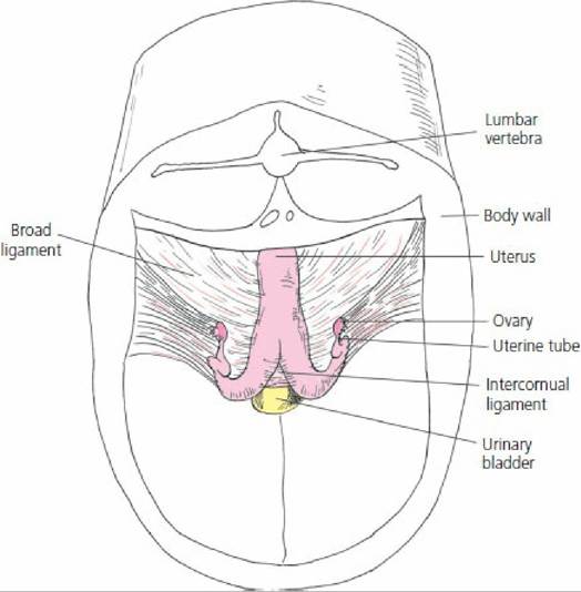
■ FIGURE 15-3 Dorsocranial view of bovine female reproductive organs. The broad ligament is the inclusive term for the mesovarium, mesosalpinx, and mesometrium, which suspend the ovary, uterine tubes, and uterus, respectively, from the dorsolateral wall of the sublumbar region.
The broad ligament is a reflection from the peritoneum.
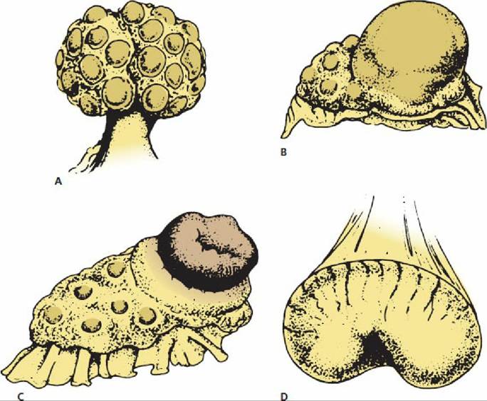
■ FIGURE 15-4 Ovarian differences resulting from species morphology and functional changes. A. Sow ovary (berry shaped). B. Cow ovary (almond shaped) with ripening follicle. C. Cow ovary with fully developed corpus luteum. D. Mare ovary (kidney shaped) with ovulation fossa (indentation on the lesser curvature). (From Dyce KM, Sack WO, Wensing CJG. Textbook of Veterinary Anatomy. 3rd edn. Philadelphia, PA: WB Saunders, 2002.)
The ovary has a surface or superficial layer of epithelium that is underlain by the tunica albuginea, a connective tissue covering the entire ovary. Beneath the tunica albuginea is the cortex, which contains a large mass of follicles in various stages of development. The medulla is centrally located and contains loose connective tissue, blood vessels, lymphatics, and nerves.
Ovarian Follicles
The follicles within the cortex are classified as: (1) primordial (sometimes called primary) follicles, (2) growing (secondary) follicles, and (3) Graafian (tertiary) follicles (Figure 15-5). The primordial follicles contain a single oocyte that is surrounded by a single layer of granulosa cells. The granulosa cells are derived from the superficial epithelium and the oocytes are derived from mitosis of oogonia in the embryonic genital ridge that then migrate to the ovary. Growing follicles are follicles that have begun growth from the resting stage as primordial follicles but have not developed a thecal layer or antrum (fluid-filled cavity; see Figure 15-5). They have two or more layers of granulosa cells surrounding the oocyte. Additional layers are added with continued growth. A zona pellucida that surrounds the oocyte may also be present. The zona pellucida provides pores through which processes of granulosa cells can interact with the oocyte surface. Also, sperm must first recognize and then contact and traverse the zona pellucida to reach the oocyte plasma membrane.
Graafian follicles are those in which an antrum is clearly visible. Two layers of thecal cells, theca interna and theca externa, are also present (see Figure 15-5).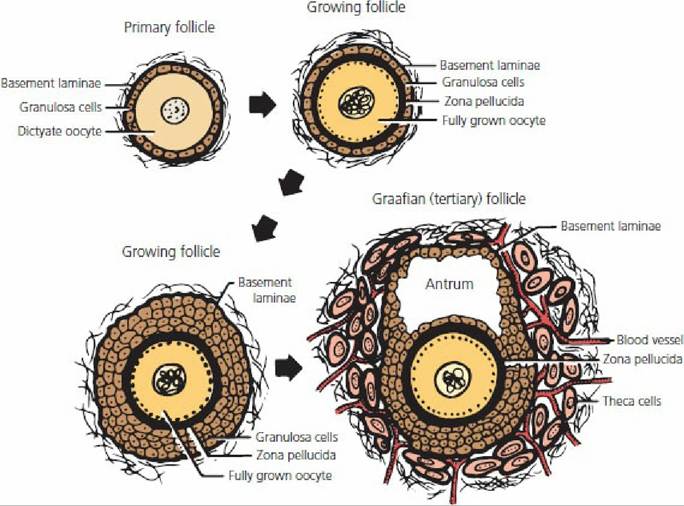
■ FIGURE 15-5 Development of an ovarian follicle from its primordial (primary) form to a Graafian follicle. Growing follicles are those that have begun growth from the resting stage as primordial follicles but have not developed thecal layers or an antrum. (From Pineda MH. Female reproductive system. In: Pineda MH, Dooley MP, eds. Veterinary Endocrinology and Reproduction. 5th edn. Ames, IA: Iowa State Press, 2003.)
Follicle Regression
Considerable atresia (regression) of the many primordial follicles occurs by birth and throughout the reproductive life of the female. At the end of the female’s reproductive life, only a few primordial follicles remain, and even these undergo atresia soon thereafter. Growth of some number of primordial follicles does occur after birth and before puberty, but these never reach the Graafian follicle stage and regress. The growth that occurs before puberty is not hormone related and is probably controlled by an unknown intraovarian factor. The formation of Graafian follicles is hormone-dependent and begins at puberty when tonic levels of LH and FSH begin to rise and fall with each estrous cycle. Many of the follicles that undergo growth and maturation with each cycle never ovulate. Therefore, the number of primordial follicles that reach the Graafian follicle stage and proceed to ovulation is a very small fraction of the birth number.
Oogenesis
The process by which oocytes are formed is known as oogenesis. The oocyte of the primordial follicle is a primary oocyte that is in a quiescent (arrested) stage of meiosis. Meiosis resumes at the time of ovulation. Whereas four spermatozoa arise from one primary spermatocyte, only one oocyte develops from the reduction division of a primary oocyte.
A polar body, which lacks sufficient cytoplasmic material for viability, develops when a primary oocyte divides to form a secondary oocyte. Another polar body is formed by the division of the secondary oocyte at the time of ovulation. The surviving oocyte has a haploid (n) number of chromosomes (similar to a spermatozoon) so that the union of a spermatozoon with an oocyte produces a cell with a diploid (2n) number of chromosomes.Tubular Genital Tract
The tubular genital tract is the location for transport of spermatozoa to the oocyte. If fertilization occurs, the tract becomes the site for development of the fetus.
Uterine Tubes
The uterine tubes are also called the oviducts and, less frequently, fallopian tubes. They are paired, convoluted tubes that conduct oocytes from the ovaries to the respective horn of the uterus. The uterine tubes serve as the site for fertilization of released oocytes by spermatozoa in domestic species. The portion of each tube adjacent to its respective ovary expands to form the infundibulum (see Figure 15-1) and fimbria project from its free edge. The fimbria assist in directing the oocyte into the infundibulum at the time of ovulation.
The lumen of the uterine tubes are lined with secretory cells and ciliated cells. These cells provide an environment for the oocytes and transport the spermatozoa. Both longitudinal and circular smooth muscles are located within the walls of the uterine tubes, which assist in the transport of oocytes and spermatozoa by their contractions. The serous covering of the uterine tubes (see Figure 15-3) is known as the mesosalpinx, which is a continuation of the mesovarium and a part of the broad ligament (providing the serous support system for the internal genitalia).
Uterus
The uterus provides a place for development of the fetus if fertilization has occurred. The uterus consists of a corpus (body), a cervix (neck), and two cornua (horns). The relative proportions of corpus, cornua, and cervix vary among species.
The corpus is largest in the mare, less extensive in the cow and sheep, and small in the sow and bitch (Figure 15-6).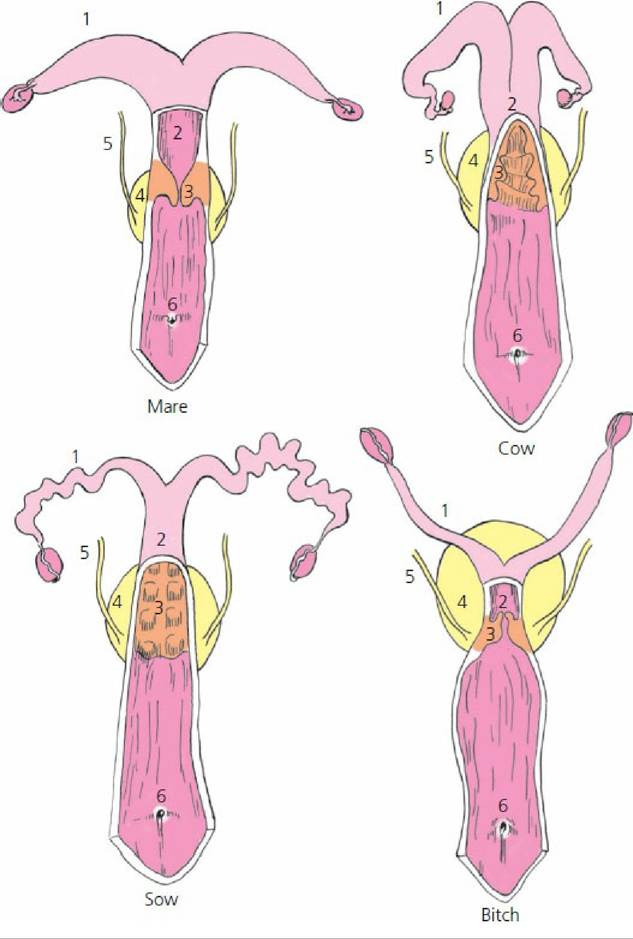
■ FIGURE 15-6 Genital tract comparisons among some domestic animals. 1, Uterine horn; 2, uterine body; 3, cervix; 4, urinary bladder; 5, ureter; 6, urethral opening. The genital tracts are opened dorsally near the body of the uterus, and the opening is extended caudally to the labia to show the cervix and urethral opening. Note that the relative proportions of uterine horns, uterine body, and cervix vary among species. The illustrations are not drawn to scale and do not compare size.
The mucous membrane lining the interior of the uterus (endometrium) is highly glandular. The glands are scattered throughout the entire endometrium of the uterus except in ruminants, in which the caruncles (mushroom-like projections from the inner surface that provide attachment for the fetal membranes) are nonglandular (Figure 15-7). The endometrium varies in thickness and vascularity with hormonal changes in the ovary and with pregnancy. The glandular secretion of the endometrium provides nutrients for the embryo before placentation (development of placental membranes), after which nutrition is provided by the mother’s blood.
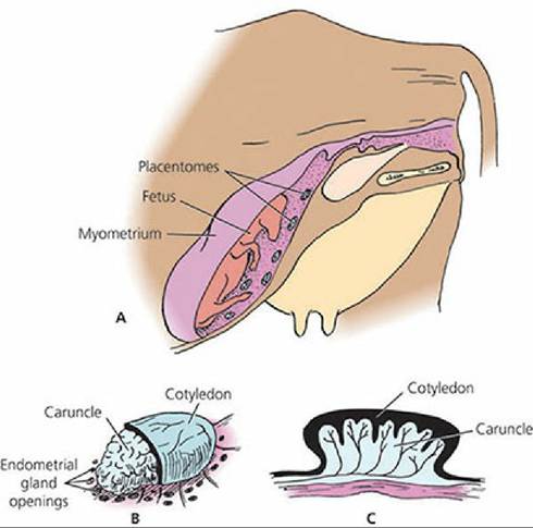
■ FIGURE 15-7 Relationship of the bovine fetal placenta to the maternal endometrium. A. View of fetus within the uterus showing multiple placentomes (caruncle and cotyledon together are referred to as a placentome). B. Magnification of a placentome that is surrounded by a number of endometrial gland openings. Only a part of the fetal cotyledon is shown so that the underlying maternal caruncle and endometrial gland openings can be visualized. C. Cross-section of a placentome. The contribution by the fetal placenta is known as the cotyledon and the maternal contribution is known as the caruncle.
The cervix projects caudally into the vagina (see Figure 15-2). This heavy, smooth muscle sphincter is tightly closed, except during estrus and at parturition (birth of young). The mucus seen at estrus is the secretion of cervical goblet cells. Goblet cell secretion of mucus during pregnancy and its outward flow prevents infective material from entering from the vagina.
The myometrium is the muscular portion of the uterus, composed of smooth muscle cells. The myometrium hypertrophies during pregnancy, increasing both in cell number and cell size. The principal function of the myometrium is aiding in the expulsion of the fetus at parturition.
The serous covering of the uterus is continuous with the mesosalpinx; in the uterus it is known as the mesometrium. The mesometrium provides a suspensory support, particularly for the nongravid uterus. It should be noted (see Figure 15-3) that there are two broad ligaments, each extending from the right or left sublumbar region and lateral pelvic wall to their respective ovary, uterine tube, and uterine horn and extending caudally on to the body of the uterus. The gravid (pregnant) uterus enlarges, and major support is provided by the abdominal wall (Figure 15-8).
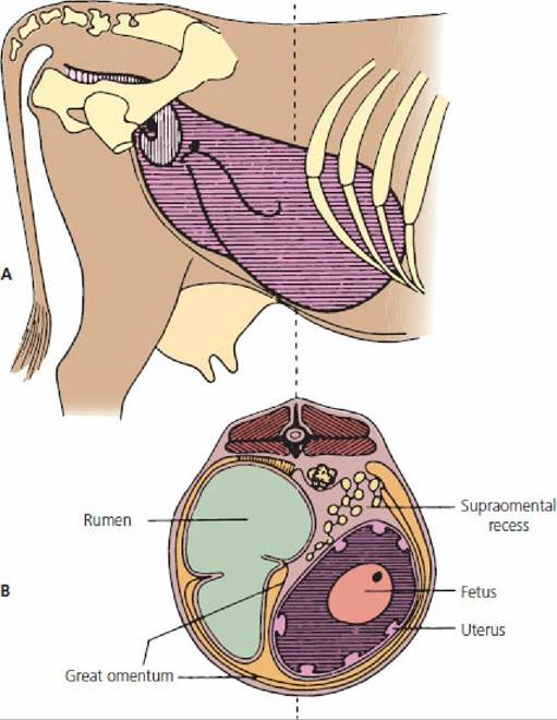
■ FIGURE 15-8 Position of the cow’s uterus. A. The nongravid uterus (vertical striping) compared with the 6-month gravid uterus (horizontal striping). B. Location of the 6-month gravid uterus in transverse section (rumen on left and uterus on right side of abdomen). (From Dyce KM, Wensing CJG. Essentials of Bovine Anatomy. Philadelphia, PA: Lea & Febiger, 1971.)
Vagina
The vagina is the portion of the birth canal located within the pelvis, between the uterus cranially and the vulva caudally (see Figures 15-1 and 15-2). The vagina serves as a sheath for the male penis during copulation. It is lined with stratified squamous epithelium, which is glandless. The fornix is the space formed cranial to the projection of the cervix into the vagina. In some animals the fornix is only visible dorsally, whereas in others it can completely encircle the cervix or be entirely absent (as in the pig).
External Genitalia
The external genitalia consists of the vulva, labia, and clitoris. The vulva is the caudal portion of the female genitalia that extends from the vagina to the exterior. The external urethral orifice (opening) is the landmark junction of the vagina and the vulva. The vestibule of the vagina (Figure 15-9) is another name for the vulva. It is the part of the external genitalia between the vagina and the labia (lips of the vulva). The clitoris (female vestigial counterpart of the penis) is concealed by the lowest part of the vulva. The clitoris is supplied with erectile tissue and sensory nerve endings. The external part of the vulva is its vertical opening, the labia (see Figure 15-1).
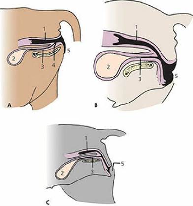
■ FIGURE 15-9 Species variations in position of the vestibule of the vagina. A. Cow. B. Mare. C. Bitch. The vulva, and hence the vestibule of the vagina, extends caudally from the external urethral orifice. 1, Vagina; 2, bladder; 3, urethra; 4, suburethral diverticulum (not present in the mare and bitch); 5, vulva. (From Dyce KM, Sack WO, Wensing CJG. Textbook of Veterinary Anatomy. 4th edn. St Louis, MO: Saunders Elsevier, 2010.)
Blood Supply of Female Genitalia
The ovary and oviduct receive their blood supply from the ovarian artery and the vagina receives its blood supply from the vaginal artery (Figure 15-10). The major blood supply to the uterus comes from the uterine artery (formerly called the middle uterine artery). The cranial part of,the uterus is also supplied with blood from the ovarian artery and the caudal part of the uterus receives blood from the vaginal artery. During pregnancy, the blood supply to the uterus increases dramatically. When the uterine artery is palpated, a vibration of the blood within it can be felt. This is called fremitus and is considered to be a good indicator of pregnancy. The ovarian artery is coiled and adheres closely to the ovarian vein, which is carrying blood from both the ovary and uterus (Figure 15-11). Such an arrangement is important for the diffusion of the hormone prostaglandin F2α (PGF2α) (see Chapter 6) from the ovarian vein to the ovarian artery in some species (e.g., cow and ewe, perhaps others). Early transport by this arrangement avoids the general circulation, where much of it would be inactivated by vascular endothelial cells in the lungs. Production requirements are lower because most of the PGF2α produced goes only to the target organ (ovary) and avoids general circulation (and subsequent inactivation) to all body parts. PGF2α at the ovarian site initiates luteolysis (termination of the corpus luteum).
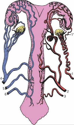
■ FIGURE 15-10 Ventral view of blood supply to the reproductive tract of the cow. The arteries are shown on the right side and the veins on the left. 1, ovarian artery; 1', uterine branch; 2, uterine artery; 3, vaginal artery; 4, ovarian vein; 5, uterine vein; 6, vaginal vein. (From Dyce KM, Sack WO, Wensing CJG. Textbook of Veterinary Anatomy. 4th edn. St Louis, MO: Saunders Elsevier, 2010.)
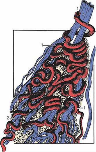
■ FIGURE 15-11 Relationship of the ovarian artery of a ruminant and its branches (1) with those of the ovarian vein (2). The intertwining ensures a large area of contact. (From Dyce KM, Sack WO, Wensing CJG. Textbook of Veterinary Anatomy. 3rd edn. Philadelphia, PA: WB Saunders, 2002.)
■