HORMONES OF FEMALE REPRODUCTION
1. Are diethylstilbestrol and estradiol-17β both estrogens? Are they both steroids?
2. Which female steroid hormone.has activities that are performed in concert with estrogens and usually requires previous estrogen priming?
3.
Which female steroid hormone prevents contractility of the uterus during pregnancy?4. What are the main functions.of the gonadotropins in the female?
5. Are tonic levels of the gonadotropins in the female increased or decreased by estrogens?
6. What is the role of the hypophysioportal system in the release of FSH and LH?
7. What is the significance of gradually increasing concentrations of estrogen over a period of time on LH release?
The principal hormones associated with ovarian cycling, pregnancy, and parturition, are estrogens, progesterone, and gonadotropins.
Estrogens
Estrogens occur naturally and synthetically. The important estrogens in mammals are steroids, produced by the ovary (granulosa cells of follicles), placenta, and adrenal cortex. A common synthetic estrogen is diethylstilbestrol, which is not a steroid but a complex alcohol with estrogenic properties. The chemical structures of diethylstilbestrol and estradiol-17β (a steroid) are compared in Figure 15-12. Regardless of production site, steroids,share a common biosynthetic pathway (Figure 15-13).
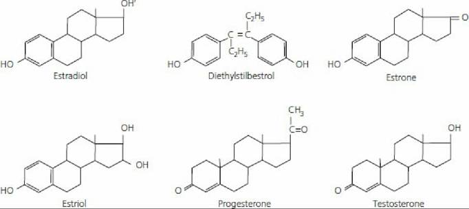
■ FIGURE 15-12 Chemical structure of some steroid hormones and diethylstilbestrol. (From Pineda MH. Female reproduction system. In: Pineda MH, Dooley MP, eds. McDonald’s Veterinary Endocrinology and Reproduction. 5th edn. Ames, IA: Iowa State Press, 2003.)
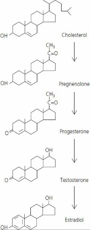
■ FIGURE 15-13 Biosynthesis of steroid hormones from cholesterol.
(From Hafez ESE, Hafez B. Reproduction in Farm Animals. 7th edn. Baltimore, MD: Lippincott Williams & Wilkins, 2000.) Estradiol-17β and estrone are estrogens that predominate in domestic nonpregnant and pregnant animals, respectively. Generally, the principal function of the estrogens is to cause cellular proliferation and growth of the tissues related to reproduction. Tissue responses caused by estrogens include:1. stimulation of endometrial gland growth;
2. stimulation of duct growth in the mammary gland;
3. increase in secretory activity of uterine ducts;
4. initiation of sexual receptivity;
5. regulation of secretion of luteinizing hormone (LH) by the anterior pituitary gland;
6. possible regulation of PGF2α release from the nongravid and gravid uterus;
7. early union of the epiphysis with the shafts of long bones, whereby growth of long bones ceases;
8. protein anabolism; and
9. epitheliotropic activity.
The protein anabolic effect of estrogens is less pronounced than that associated with testosterone. Its effect is probably associated more specifically with the sex organs rather than with a generalized effect. The epitheliotropic function manifests at estrus when the epithelium in the vagina proliferates and cornification is more prevalent.
Progesterone Progesterone, like the estrogens, is a steroid sex hormone produced by the corpus luteum (CL) of the ovary, placenta, and adrenal cortex. Its place in the common biosynthetic pathway scheme is shown in Figure 15-13. It is the principal progestational hormone. Certain synthetic and natural progestational agents are called progestins.
The activities associated with progesterone are often performed in concert with estrogens, and usually require previous estrogen priming. The functions of progesterone include: (1) promotion of endometrial gland growth, (2) stimulation of secretory activity of the oviduct and endometrial glands to provide nutrients for the developing embryo before implantation, (3) promotion of lobuloalveolar growth in the mammary gland, (4) prevention of contractility of the uterus during pregnancy, and (5) regulation of secretion of gonadotropins.
The interrelationships of the estrogens, progesterone, and gonadotropins are described later in the discussions of the estrous cycle and pregnancy.
Gonadotropins
Follicle-stimulating hormone (FSH) and luteinizing hormone (LH) are collectively referred to as the gonadotropins because of their role in stimulating cells within the ovary and testis (the gonads). FSH and LH are hormones secreted by cells within the anterior pituitary. Both are classified chemically as glycoproteins. A glycoprotein is a conjugated protein in which the nonprotein group is a carbohydrate.
The main function of FSH in the female is promotion of the growth of follicles. LH is important for the ovulatory process and the luteinization of the granulosa, an essential aspect of CL formation. Apparently, FSH and LH concentrations exist in the plasma at a tonic or basal level. These levels are controlled by negative feedback from the gonads. Tonic levels are increased by estrogen and decreased by progesterone.
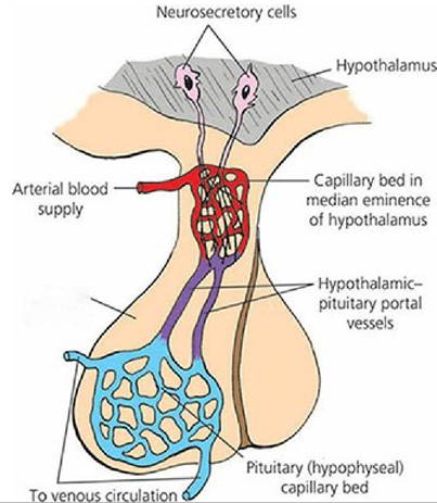
The release of FSH and LH from the anterior pituitary is controlled by a releasing hormone from the hypothalamus. The circulatory system involved is known as the hypophysioportal system (Figure 15-14). A portal system begins with capillaries and terminates with capillaries. The hypothalamic capillaries receive a secretion from sensing cells in the hypothalamus known as gonadotropin releasing hormone (GnRH). GnRH is secreted in response to low levels of LH or FSH and is then followed by secretion of LH or FSH.
Anterior pituitary
■ FIGURE 15-14 The hypophysioportal circulation involved with the secretion of anterior pituitary hormones. Cell bodies in the hypothalamus sense the need for a hormone and secrete a releasing hormone into the hypothalamic capillary bed. The releasing hormone enters the hypophyseal capillary bed and diffuses to specific cells, causing them to secrete their specific hormone.
The concentrations of estrogens and progesterone also influence the amount of LH or FSH secretion. Generally, an increasing concentration of estrogen causes an increase in sensitivity of the anterior pituitary to GnRH, and results in an increased release of gonadotropins. Progesterone decreases sensitivity of the anterior pituitary to GnRH, and LH and FSH concentrations decrease. These influences, particularly those of estrogen, depend on gradually increasing concentrations of estrogen over a period of time, which results in the preovulatory surge of LH release. Conversely, when estrogen concentration is basal and of short duration, LH and FSH secretions are suppressed.