FUNCTIONAL ASPECTS
Gilts attain puberty around 6 months. The species is polyestrus: the cycle repeats at intervals of about 21 days. Fertilization takes place in the ampullae, where the conceptuses are detained for a few days before being admitted to the uterus.
Cleavage continues there, creating blastocysts that are initially spherical and randomly placed. By the end of 2 weeks they have become filamentous and greatly lengthened—up to 60 cm—and have adopted permanent, regularly spaced stations that make full use of both horns, which is an arrangement that may have required some conceptuses to migrate from one horn to the other. The conception rate is high, but so also is prenatal mortality—40% or more. The placenta is of the diffuse epitheliochorial type. Antibody transfer does not occur in utero, and the newborn is dependent on the ingestion of colostrum for its initial immunological protection.During pregnancy, the horns increase greatly in diameter, and their length may double. Growth of the tissues within the broad ligaments allows the horns to sink into the ventral half of the abdomen, where they push the intestines craniodorsally and make contact with the stomach and liver; they carry the ovaries with them, taking them out of reach of a hand within the rectum. Confirmation of pregnancy at this stage is provided by the firmness of the cervix and, more reliably, by the characteristic fremitus of the enlarged uterine artery. An alternative, less troublesome method of pregnancy diagnosis is available in ultrasonography, with the use of either a transabdominal or transrectal approach (Figure 35-4, B-C).
Gestation lasts 114 days (on average), and farrowing is preceded by the usual relaxation of the joints and
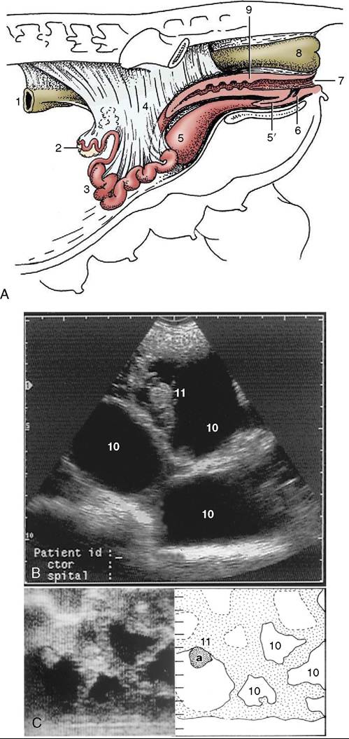
Figure 35-4 A, The reproductive organs of the sow in situ.
(The presence of the intestines in the intact animal causes the ovaries and uterine horns to lie more dorsally than shown here.) Transrectal (B) and transabdominal (C) ultrasonographic images of 30-day gravid porcine uteruses. (Scales in centimeters.) 1, Descending colon; 2, ovary; 3, uterine horns; 4, broad ligament; 5, bladder; 5', urethra; 6, suburethral diverticulum; 7, vulva; 8, rectum; 9, cervix; 10, allantoic fluid-filled spaces; 11, (a), embryo.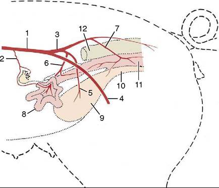
Figure 35-5 The principal arteries supplying the left side of the female reproductive tract, schematic. 1, Aorta; 2, ovarian a. with cranial uterine branch; 3, internal iliac a.; 4, external iliac a. continued by femoral into left thigh; 5, umbilical a.; 6, left uterine a. crossing medial surface of external iliac; 7, vaginal a. with caudal uterine branch; 8, left uterine horn; 9, bladder; 10, urethra; 11, vagina; 12, rectum.
From Evans HE, Sack WO: Prenatal development of domestic and laboratory animals. Growth curves, external features, and selected references. Anat Histol Embryol 2:11-45, 1973.