Ganglion CellAxonsTransmitAction Potentials to the Visual Cortex by Way of the Lateral Geniculate Nucleus
Sets of retinal ganglion cells leaving the eye participate in three important visual pathways: the retino-genieulo- Striate pathway, the retino-tectal pathway, and the retino- Iiypothalaniic pathway.
The retino-geniculo-striate pathway is principally involved in conscious visual perception of form, color, motion, orientation, and depth. The retino-tectal pathway plays an important role in pupillary reflexes and reflex orientation of the eye to visual targets. The retino- hypothalamic pathway plays a role in the regulation of physiological rhythms by light-dark cycles (e.g., seasonal changes in day length).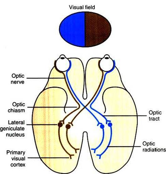
FIGURE 14-7 Retino-geniculo-striate pathway to cerebral cortex. Axons of the retinal ganglion cells project to the lateral geniculate nucleus of the thalamus, and axons from cells of the lateral geniculate nucleus project to the ipsilateral primary visual cortex of the occipital lobe.The right environmental visual field projects to the left half of each retina, and the left environmental visual field projects to the right half of each retina.The right half of each retina sends information to the right primary visual cortex, and the left half of each retina sends information to the left primary visual cortex.Therefore, information from the left visual field arrives in the right primary visual cortex, and information from the right visual field arrives in the left primary visual cortex.The labeled components of each pathway are present on both sides of the brain.
In order for the retinal image, originating from light in the visual field, to reach consciousness, the information must be transferred to the visual cortex. Figure 14-7 shows the retino- geniculo-striate pathway by which the axons of the retinal ganglion cells project to the lateral geniculate nucleus of the thalamus and by which cells in the lateral geniculate nucleus project their axons to the primary visual cortex of the occipital lobe.
Note that ganglion cell axons from the temporal retina (closest to the ear; see Figure 14-1) travel along the optic nerve to the optic chiasm and then project ipsilaterally to the lateral geniculate nucleus on the same side of the brain. Ganglion cell axons from the nasal retina (closest to the nose) come to the optic chiasm and cross to the contralateral lateral geniculate nucleus. Retinal ganglion cell axons between the region of the optic chiasm and the lateral geniculate nuclei are referred to as the optic tracts. Cells in each lateral geniculate nucleus then send axons to the ipsilateral primary visual cortex, in the occipital lobe, by way of the optic radiations. This arrangement results in the right half of each retina sending information to the right primary visual cortex and the left half of each retina sending information to the left primary visual cortex. Light originating in the left half of the environmental visual field generally falls on the right half of the retina of each eye, whereas light originating in the right half of the environmental visual field falls on the left half of the retina of each eye. Given the anatomical map of the retino-geniculo-striate pathway in Figure 14-7, information from an image arising in the left field of vision would be received in the right primary visual cortex, and image information arising from the right field of vision would be received in the left primary visual cortex.Information reaching primary visual cortex, or Vl (also called striate cortex), is passed to other occipital lobe areas of visual cortex (e.g., V2, V3) for more complex processing, then to visual association areas of the parietal and temporal lobes for the highest forms of visual information processing and integration with other sensory modalities. There appears to be parallel processing of retinal information within the retino- geniculo-striate pathway and its higher-order processing regions. One major stream of information in this system apparently is concerned with conscious object identification (e.g., form, color), whereas a parallel stream of information relates to conscious awareness of where an object is located (e.g., motion, position). The three major projection pathways of retinal ganglion cell axons—retino-geniculo-striate, retino- tectal, and retino-hypothalamic—represent another example of the parallel processing of retinal information.