Gastric Physiology
Gastric Glands and Secretions
The term gastric juice refers to the combination of substances secreted into the stomach lumen by gastric glands, also termed gastric pits because of their pitlike extension into the wall of the stomach (Fig.
21-3), and epithelial cells of the stomach mucosa. Gastric juice contains water, hydrochloric acid, mucus, intrinsic factor, pepsinogen (an inactive form of pepsin, a proteolytic enzyme), and the enzyme rennin. The regulation of gastric juice secretion has three phases, cephalic, gastric, and intestinal.stimulation of gastric secretions during the cephalic phase is in response to the sight, smell, or taste of food. These induce a neural response
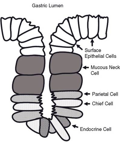
Figure 21-3. Gastric pit (gastric gland) in the lining of the stomach. Note the different cell types found in the epithelium lining the gland.
that increases parasympathetic (vagal nerve) stimulation to the stomach, and this stimulates gastric secretions. The gastric phase begins when food enters the stomach.
The presence of food, especially proteins, stimulates the secretion of the hormones gastrin and histamine from cells in the gastric epithelium. Gastrin and histamine stimulate parietal cells in gastric glands to secrete hydrochloric acid (Fig. 21-3). Acetylcholine (parasympathetic neurotransmitter) also stimulates parietal cells to secrete hydrochloric acid, but all three regulators (gastrin, histamine, and acetylcholine) must be present for the most efficient hydrochloric acid secretion. The histamine receptors on parietal cells (H2 receptors) are different from those on cells involved in allergic reactions (H1 receptors). The specific H2 receptor antagonists provide a means to reduce acid secretion with few side effects.
The antihistamines used for allergies do not bind to H2 receptors and thus do not disturb digestion. The hormones cholecystokinin, gastric inhibitory peptide, and secretin inhibit hydrochloric acid secretion. These hormones are released from the duodenal epithelium in response to the presence of food in the duodenum. The release of these hormones that act to inhibit gastric function is part of the intestinal phase of gastric regulation.The pH of gastric juice in mammals can be 2 or less. The low pH is protective in that most foreign microbes ingested with food cannot survive such an acidic environment. The low pH inhibits hydrochloric acid secretion to prevent it from becoming too acidic. Pepsinogen (an inactive form of the enzyme pepsin and a component of gastric juice) is activated by the low pH. By its proteolytic activity, pepsin can activate more pepsinogen. The low pH also promotes the activity of pepsin, because the most favorable pH range for its proteolytic activity is 1.3 to 5. Chief or peptic cells (Fig. 21-3) secrete pepsinogen to begin protein digestion in the stomach, but protein digestion is completed in the small intestine by other digestive enzymes.
A layer of mucus covers the epithelial lining of the stomach and protects the epithelium from the low pH of the gastric fluids. This mucus is produced by cells in the gastric glands (Fig. 21-3) and is secreted from there onto the surface of the epithelium. Mucus secretion is stimulated by prostaglandins, which are also produced locally in the wall of the stomach. Non-steroidal anti-inflammatory drugs (such as aspirin and phenylbutazone) inhibit the synthesis of prostaglandins, and toxic doses of these agents are associated with gastric ulcers. It is presumed that a lack of mucus secretion contributes to the development of the ulcers.
Rennin is an enzyme in the gastric juice in the abomasum of young ruminants. its function is to coagulate milk and reduce its rate of passage through the gastrointestinal tract.
intrinsic factor, a carrier protein for vitamin B12, binds to the vitamin, and the resultant complex passes through the tract to the ileum, which absorbs the B12.Gastric Motility
Gastric movements mix the ingesta with the gastric juice, continue mechanical digestion (to liquefy the digesta), and pass the digesta into the duodenum at a controlled rate. The stomach regularly produces peristaltic contractions, beginning in the region of the cardia and increasing in force as they travel over the stomach to the pyloric antrum (see Fig. 20-8). These mix and grind the food and force some through the pyloric sphincter into the duodenum. However, much of the food (and especially larger particles) is held back to allow for more mixing and grinding. The ingesta forced through the pyloric sphincter, termed chyme, is a mushy, semisolid mixture of food, water, and gastric juice.
similar to the regulation of gastric secretions, the regulation of gastric motility can be divided into cephalic, gastric, and intestinal phases. stimulation during cephalic regulation occurs via the parasympathetic nerves, and this increases in response to sight, smell, or taste of food. The hormone gastrin stimulates overall gastric motility to promote mixing (gastric phase). The hormones cholecystokinin and secretin and gastric inhibitory peptides promote a more forceful contraction of the pyloric sphincter to slow gastric emptying (intestinal phase). The inhibitory effect of the duodenal hormones (released in response to chyme entering the duodenum) prevents the delivery of chyme to the duodenum too fast to be digested normally.
The stomach of a carnivore empties within a few hours, usually before the next meal. Other animals require many hours to empty the stomach. Both the horse and pig require a full day’s fast (24 hours) to empty a full stomach. The stomach of a nursing foal empties slowly, but in an adult pony liquid passes from the stomach to the cecum in 2 hours.
in addition to the typical pattern of stomach contraction when food is present, waves of peristaltic contractions may occur over the stomach as a slight ripple. These are produced by spontaneous electrical depolarizations, which sometimes induce action potentials, in the smooth muscle. These begin in the cardia region, and the waves of membrane depolarization are termed gastric slow waves. In prolonged fasting, the magnitude of the contractions becomes greater (hunger contractions). These are apparently a response to an increase in parasympathetic input during prolonged fasting. These reach maximum intensity in humans after about 3 days without food and weaken progressively thereafter. In the horse, hunger contractions may begin as early as 5 hours after eating, when the stomach still contains some food. The intensity of hunger contractions is related to the level of blood sugar. As the blood sugar level decreases, the intensity of hunger contractions increases.