GENERAL ANATOMY OF THE PELVIS AND PERINEUM (See also pp. 55-56.)
The bony pelvis is formed by the pelvic girdle, sacrum, and first few caudal vertebrae; of course the caudal limit of the roof is, as always, difficult to define precisely. The bones were described in Chapter 2, and the surface landmarks they create are mentioned in Chapter 17.
It will therefore be sufficient at this point to recapitulate a few general features of the anatomy of the pelvis.The pelvic cavity is smaller than might be supposed from examination of the intact animal or the isolated girdle. The discrepancy between expectation and reality is due to the shallowness of the caudal part of the abdomen and to the acute angle (about 20°) formed between the ilia and the vertebral column (Figure 15-1, A-B). The pronounced obliquity of the inlet places the pubic brim level with, or even behind, the caudal limit of the sacrum. The iliac shafts are not quite parallel, and the inlet is widest in its middle part and narrowest dorsally. The pelvic outlet is less confined than the inlet and possesses a considerable capacity for further enlargement through elevation of the tail behind the very short sacrum. Only a small part of the lateral wall is bony, as neither the ischial spine nor the ischial tuber rises to any great height. In the dog the sacrotuberous ligament is reduced to a narrow cord (under cover of the superficial gluteal muscle) extending between the ischial tuber and the caudolateral corner of the sacrum (Figure 15-1, A).
The pelvic girdle of the cat shows some differences. Cranially, the ilia diverge slightly, producing a somewhat funnel-shaped entrance to the pelvis from the abdominal cavity. The wings of these bones are relatively smaller and shallower, which also eases the transition. The ischial tubers stand closer together than in the dog, which gives the pelvis a more rectangular appearance in the ventrodorsal view and a more confined exit (Figure 15-2).
In consequence of the last feature the perineum is narrow. There are no sacrotuberous ligaments in this species.The axis of the short pelvic canal is almost straight, and in general the conformation appears well adapted for easy parturition. Sexual dimorphism is not pronounced, and pelvic measurements have not been given much attention in small animal obstetrics. An ill match of the proportions of the fetus and the dam is most common in cases in which the litter is small (and the individual fetus relatively large) in toy dogs, and in those breeds in which a measure of achondroplasia is a feature of the conformation. On rectal examination the pelvic canal of young dogs is shaped like an hourglass, which may mistakenly suggest a pelvic fracture.
The perineum slopes somewhat ventrocaudally and is largely concealed when the tail is carried low. When the tail is raised, it exhibits a shield of naked integument about the anal orifice and, at some distance ventral to this, the vulva or root of the penis; these features are considered in more detail later. The ischiorectal fossa between the anus and the ischial tuber naturally varies in prominence with the character of the coat and the degree of obesity. The fossa is bounded by the sacrotu- berous ligament and the deep face of the superficial gluteal muscle laterally and by the superficial face of the coccygeus medially. It is traversed by the large caudal gluteal vessels that run against the lateral wall and by the main trunks and certain branches of the internal pudendal vessels and pudendal nerve placed more medially, toward the floor (Figure 15-17/2,3).
The pelvic diaphragm has the usual composition. The lateral muscle, the coccygeus, has a tendinous origin from the ischial spine and inserts on the lateral aspect of the tail between the second and fifth vertebrae (Figure 3-48 and Figure 15-17). The deeper and thinner levator ani (Figure 3-48/2) has a wider origin, which extends from the iliac shaft onto the pelvic floor along which it runs, directly to the side of the symphysis (Figure 15-3/7).
The part arising from the pelvic floor closely embraces the pelvic viscera in its passage to its insertion on the tail, reaching as far caudally as the seventh vertebra. The levator fibers run more obliquely than those of the coccygeus, and part of the levator emerges superficially behind that muscle. The levator has only a passing fascial connection with the external sphincter of the anus, and like the coccygeus, it is primarily a depressor of the tail. However, its fascial attachment enables it to help fix the position of the anus during defecation. The tone of both muscles is important in retaining the pelvic viscera in place, and perineal hernia—in which pelvic organs are displaced to form a swelling to the side of the anus—may be a sequel to their paralysis or atrophy. Surgical repair of this condition involves suture of the external sphincter to the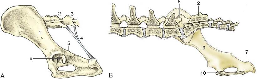
Figure 15-1 A, Canine sacrotuberous ligament, left lateral view. B, The right half of the canine bony pelvis, medial view.
1, Ilium; 2, sacrum; 3, caudal vertebra(e); 4, sacrotuberous ligament; 5, ischial spine; 6, acetabulum; 7, ischial tuber; 8, sacroiliac joint; 9, shaft of ilium; 10, symphysis.
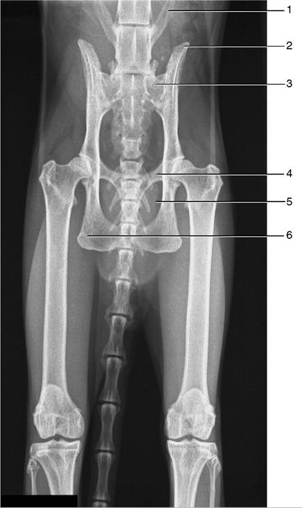
Figure 15-2 Radiograph of the feline pelvis. 1, Transverse process of last lumbar vertebra (L7); 2, iliac crest; 3, sacrum; 4, pecten of the pubis; 5, obturator foramen; 6, ischial tuber.
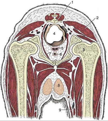
Figure 15-3 Transverse section of the canine pelvis at the level of the hip joint. 1, Caudal vertebra; 2, superficial gluteal muscle; 3, head of femur in acetabulum; 4, rectum suspended by a short mesorectum; 5, vagina; 6, urethra; 7, levator ani; 8, inguinal mammary gland; 9, femoral artery and vein.
coccygeus, internal obturator, and sacrotuberous ligament about the margins of the space.
The pelvic blood vessels and nerves were sufficiently described in the general accounts (pp. 251 and 325). Because there are only three sacral spinal nerves, the origins of the pudendal, caudal rectal, and pelvic nerves are rather compressed; variations in the branching patterns of the first two are common. The pudendal and caudal rectal nerves supply afferent and efferent fibers to the perineum, and their integrity is necessary for the execution of the perineal reflex that provides a means of gauging the depth of narcosis. The modified skin about the anus is especially sensitive, and even a gentle touch evokes a brisk contraction of the anal sphincter of the conscious or lightly anesthetized animal.