GENERAL EXTERNAL ANATOMY
There are four main fur types. These are based on fur length and range from the Rex and Satin at 12 mm, to normal rabbits at 30 mm and the Angora rabbit at as long as 120 mm. Normal rabbits have stiff, long guard hairs with a soft underfur while Rex and Satin breeds have guard hairs as short, or shorter, than the undercoat.
There are hairless areas on the nose, part of the scrotum, and in the inguinal areas in both sexes. Rabbits do not have footpads and the toes and metatarsals are covered in coarse fur. The rabbit stands plantigrade, with the whole area from hock to toe in contact with the ground, but becomes digitigrade when running (Fig. 8.2).
There is a well-developed third eyelid, which covers the eye during anesthesia or sleep. In low light the pupils dilate widely to allow for increased retinal sensitivity (Cruise & Nathan 1994). Rabbit eyes readily detect motion and are particularly sensitive to blues and greens at twilight (Harkness & Wagner 1995). The mouth is very small. The upper lip has a median cleft or philtrum that curves to the right and left around the nose - hence the term “harelip”. The neck has a pendulous skin fold called the dewlap, which is more pronounced in the doe and certain breeds (Fig. 8.2).
Head and neck
CLINICAL NOTE
The rabbit has long funnel-like ears and the pinna is composed of a thin layer of skin overlying elastic cartilage. It is freely movable and capable of independent action. Inside the ear canal a cartilaginous ridge, the tragus, separates the ear canal from a blind-ended diverticulum. Sebaceous glands are present in the skin. The pinnae represent 12% of the rabbit's body surface and so are a major site for heat loss and gain (Brewer & Cruise 1994; Cruise & Nathan 1994). There is a highly visible central artery and peripheral veins, which form large arteriovenous shunts when heated (Fig.
8.1).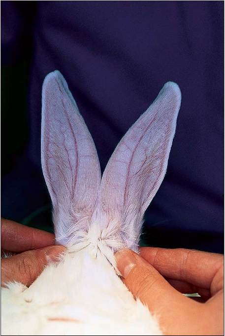
Figure 8.1 • The rabbit ears represent 12% of surface area so are a major source of heat exchange. There is a highly visible central artery and peripheral veins which form large arteriovenous shunts when heated.
The dewlap is often a site of wet dermatitis. This is caused by increased salivation due to dental pain, continual wetting of the skin from water bowls and poor hygiene of bedding. Obese animals, or does with large dewlaps, are more likely to suffer from this problem (Cruise & Nathan 1994).
External genitalia
The male has a rounded penile sheath and a round urethra with oblong scrotal sacs on each side. The penis can easily be extruded. The female vulva appears triangular and has a slitlike orifice (Fig. 8.3). The mammary glands (usually eight in number) are located along the thoracic and inguinal region. Only the female has nipples (Cruise & Nathan 1994). In the perineal region of both sexes there are two hairless patches lateral to the anus that contain the waxy inguinal glands.