SKELETAL SYSTEM
The skeleton of the pet rabbit is fragile in comparison with its heavy musculature (Fig. 8.4). For example, in the New Zealand White rabbit the skeleton accounts for 6%, while the muscles total 56% of body mass (Jelenko et al.
1971).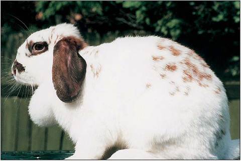
Figure 8.2 • Rabbit doe showing the dewlap, which is prone to wet dermatitis.
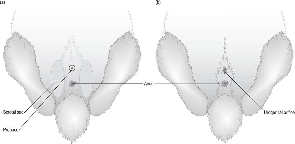
Figure 8.3 • External genitalia.
(a) Male - the urethra is round and the penis can be extruded.
(b) Female - the vulva is triangular with a slitlike orifice.
If the rabbit's spine is unsupported while being handled the heavy hindquarters twist about the Iumbrosacral junction to cause fractures (Harkness & Wagner 1995). L6-7 is a common site for spinal fractures, especially in young rabbits.
The vertebral formula is C7, T12-13, L7, S4, C15-16 (Cruise & Nathan 1994). The costal cartilages of the first seven ribs (true ribs) articulate with the sternum. The 7th to 9th ribs have attached costal cartilages while the 10th to 12th ribs are floating. There are seven sternebrae. The transverse processes of the lumbar vertebrae are long and narrow (Okerman 1994).
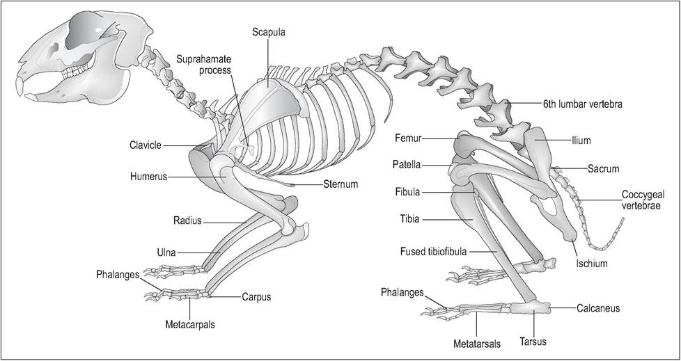
Figure 8.4 • Skeleton of the rabbit (Oryctolagus cuniculus). From Popesko, P., Rajtova, V., & Horak, J. (1990) A colour atlas of anatomy of small laboratory animals. Vol. 1. Aylesbury, UK: Wolfe with permission.
The pectoral girdle contains the scapulae and small paired clavicles. The scapula has a much more triangular infraspinous fossa than that in the cat, which is more rounded. The acromion process has a bony projection jutting at right angles, which is called the superhamate process (Fig. 8.5) (Okerman 1994).
The carpus has two rows: four bones proximally and five distally (Fig. 8.6). There are five digits and each has three phalanges, except for the first digit, which has a shorter metacarpal and only two phalanges (Cruise & Nathan 1994).The rabbit has a small accessory bone called the os acetabulum, which helps form the acetabulum, along with the ischium and ilium (Cruise & Nathan 1994). The obdurator foramina are oval in shape. The femur articulates only with the tibia; the thin, blade-like fibula is fused with the tibia for over half its length (Cruise & Nathan 1994; Okerman 1994)(Fig. 8.7). There are six tarsal bones arranged in three rows: the proximal row contains the two large bones, the tarsus and calcaneus; the middle row has one central bone, and the distal row has three bones (the 2nd, 3rd, and 4th tarsal bones) (Fig. 8.8). Metatarsals 2-5 are well developed, with a rudimentary metatarsal 1. The
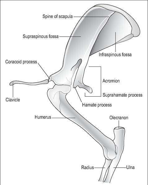
Figure 8.5 • Left shoulder of the rabbit - the acromion process has the bony superhamate process jutting at right angles. The infraspinous fossa is more triangular than in the cat. From Popesko, P., Rajtova, V., & Horak, J. (1990) A colour atlas of anatomy of small laboratory animals. Vol. 1. Aylesbury, UK: Wolfe with permission.
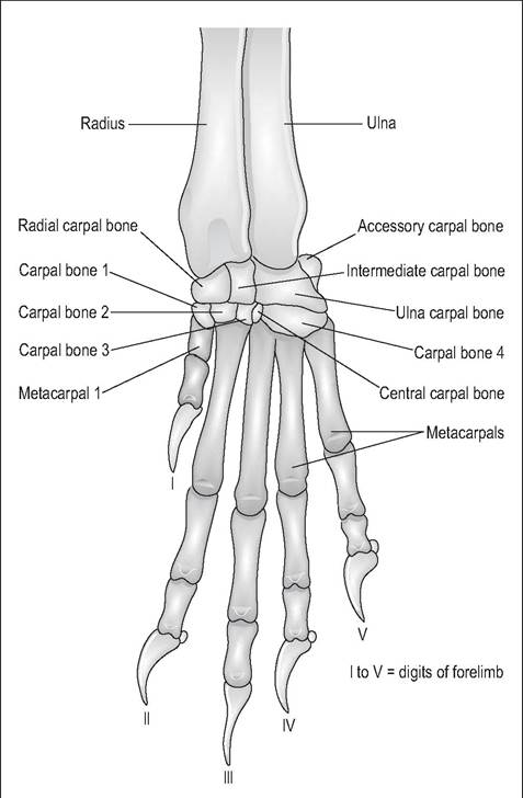
Figure 8.6 • Dorsal view of left carpus of the rabbit. From Popesko, P., Rajtova, V., & Horak, J. (1990) A colour atlas of anatomy of small laboratory animals. Vol. 1. Aylesbury, UK: Wolfe with permission.
four digits each have three phalanges (Cruise & Nathan 1994).