GENERAL FEATURES OF THE SKELETON
1. Differentiate between the axial and the appendicular skeleton.
2. What are the components of the axial skeleton?
3. What is a collective term for the bones of the head?
4.
What are the group names for the vertebrae, named in order, beginning with those most cranial?5. What is an intervertebral disk?
6. What constitutes a prolapsed intervertebral disk?
7. What is the prominent bone in the pectoral girdle of domestic mammals?
8. What bones comprise the os coxae? Know the orientation of these bones to each other.
9. Note the location of the obturator foramen.
0. Name the bones in the hindlimb.of the horse that are distal to the hock.
11. What are sesamoid bones?
2. What is the coffin joint?
The bones of the body are generally similar among the animals but vary according to size, shape, and number. The skeletons of the horse (Figure 7-1), the ox (Figure 7-2), and the fowl (Figure 7-3) are shown as examples that feature the general arrangement of the bones and their similarities. The bones of the skeleton are classified as belonging to either the axial skeleton or the appendicular skeleton.
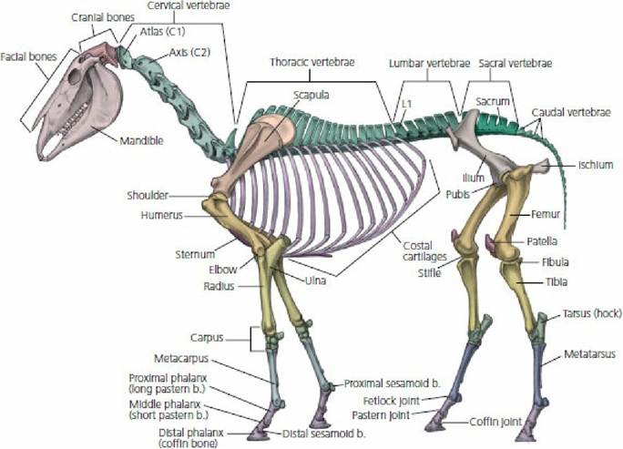
■ FIGURE 7-1 Skeleton of the horse. (Adapted from McCracken TO, Kainer RA, Spurgeon TL. Spurgeon’s Color Atlas of Large Animal Anatomy: The Essentials. Baltimore, MD: Lippincott Williams & Wilkins, 1999.)
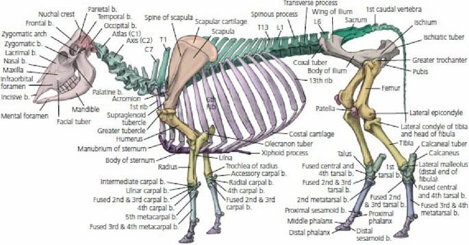
■ FIGURE 7-2 Skeleton of the ox. (From McCracken TO, Kainer RA, Spurgeon TL. Spurgeon’s Color Atlas of Large Animal Anatomy: The Essentials. Baltimore, MD: Lippincott Williams & Wilkins, 1999.)
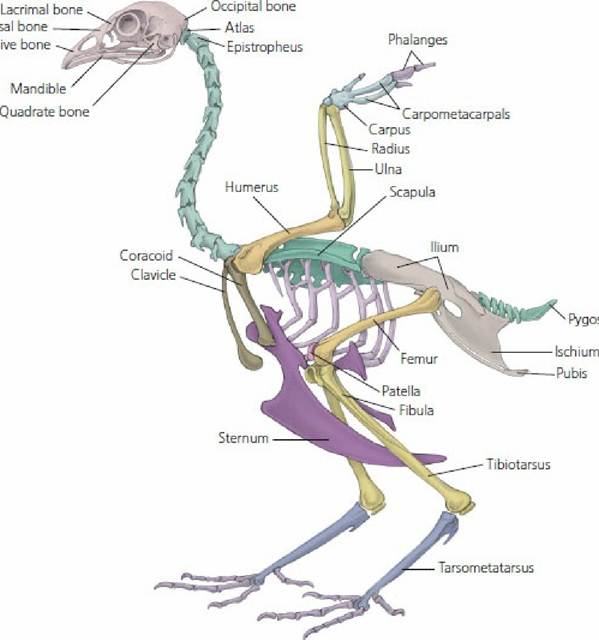
■ FIGURE 7-3 Skeleton of the chicken.
(Adapted from McCracken TO, Kainer RA, Spurgeon TL. Spurgeon’s Color Atlas of Large Animal Anatomy: The Essentials. Baltimore, MD: Lippincott Williams & Wilkins, 1999.)The Axial Skeleton
The components of the axial skeleton lie on the long axis (midline) of the body and include the skull, vertebrae, and those bones attached to the vertebrae, the ribs, and the ventral connections of the ribs, the sternum.
The skull comprises the neurocranium (brain case) and the viscerocranium (bones of the face). Cranium is a collective term for the bones of the head. The brain case provides protection for the brain and openings for cranial nerve connections. The bones of the face provide a location and
protection for the organs of the special senses and openings for the digestive and respiratory systems. Special features are considered in respective chapters.
The ribs and sternum give limits and provide protection for the thoracic viscera (heart and lungs) and, because of their movement potential, assist respiration and blood flow.
General features of vertebrae are shown in Figure 7-4. More specific information for the dog was presented when describing the relationship of vertebrae to spinal nerves (see Chapter 4). The numbers associated with the regions of their location, cervical (C), thoracic (T), lumbar (L), sacral (S), and caudal (Cd), are represented by a vertebral formula for each species. The vertebral formula for the horse is C7, T18, L6, S5, Cd15-20. The numbers of vertebrae for each region for the domestic animal species and human are presented in Table 7-1.
| TABLE 7-1 VERTEBRAL FORMULAS OF COMMON DOMESTIC ANIMALS AND HUMANS | |||||
| SPECIES | CERVICAL | THORACIC | LUMBAR | SACRAL | CAUDAL |
| Horse | 7 | 18 | 6 | 5 | 15-20 |
| Ox | 7 | 13 | 6 | 5 | 18-20 |
| Sheep | 7 | 13 | 6-7 | 4 | 16-18 |
| Goat | 7 | 13 | 7 | 4 | 12 |
| Hog | 7 | 14-15 | 6-7 | 4 | 20-23 |
| Dog | 7 | 13 | 7 | 3 | 20-23 |
| Chicken | 14 | 7 | 14 (lumbosacral) | 6 | |
| Human | 7 | 12 | 5 | 5 | 4 |
| From Frandson RD, Wilke WL, Fails AD. Anatomy and Physiology of Farm Animals. 7th edn. Ames, IA:.Wiley-Blackwell, 2009. | |||||
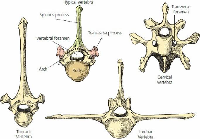
■ FIGURE 7-4 General features of typical vertebrae. (From Frandson RD, Wilke WL, Fails AD. Anatomy and Physiology of Farm Animals. 7th edn. Ames, IA: Wiley-Blackwell, 2009.)
The bodies of contiguous vertebrae are held together by a modified symphysis, which is a slightly movable joint where bones are held together by a combination of hyaline cartilage and fibrocartilage. A true symphysis consists only of Iibrocartilage. For the vertebrae, the modified symphysis is known as an intervertebral disk (Figure 7-5). The cranial and caudal surfaces of contiguous vertebrae have a covering of hyaline cartilage, and the interconnection of these coverings is the intervertebral disk. The soft gelatinous interior of the disk is termed the nucleus pulposus. The fibrocartilaginous collar that supports the periphery of the disk is termed the annulus fibrosus. The intervertebral disk (nucleus pulposus and annulus fibrosus) provides for a compression-resisting cushion that permits limited movement between contiguous vertebrae. A prolapsed or herniated intervertebral disk occurs when the nucleus pulposus herniates (ruptures) through the annulus fibrosus. When inflammation proceeds at the site of herniation, there may be spinal nerve root compression at that level and peripheral nerve involvement with loss of function for the region served.
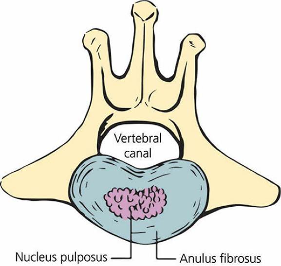
■ FIGURE 7-5 Intervertebral disk, which is composed of hyaline cartilage and fibrocartilage and serves,to interconnect the bodies of contiguous vertebrae. The soft gelatinous interior of the disk is the nucleus.pulposus. The fibrocartilagenous collar supports the periphery of the disk. A prolapsed disk occurs when the nucleus pulposus herniates through the annulus fibrosus.
(From Uemura EE. Fundamentals of Canine Neuroanatomy and Neurophysiology. 1st edn. Ames, IA: Wiley-Blackwell, 2016.)The Appendicular Skeleton
The appendicular skeleton is made up of the bones of the front (thoracic) and hind (pelvic) limbs and their respective pectoral girdle (shoulder) and pelvic girdle (pelvis). The pectoral girdle is composed of the scapula, clavicle, and coracoid, and the pelvic girdle is composed of the ilium, ischium, and pubis.
Whereas the pectoral girdle of birds has a visibly discernible scapula, coracoid, and clavicle, the only bone of prominence in the pectoral girdle of domestic mammals is the scapula (shoulder blade). The coracoid is reduced to a small coracoid process projecting medially from the supraglenoid tubercle, from which the coracobrachialis muscle arises. This muscle stabilizes the shoulder. The clavicle is represented only by a fibrous identity in the brachiocephalicus muscle (a muscle of the forelimb) except in the cat, in which a very small clavicular bone is embedded in this muscle. It has no function but will appear on radiographs, where it may be erroneously interpreted as a bone lodged in the esophagus. Medial and lateral views of the horse scapula are shown in Figure 7-6.
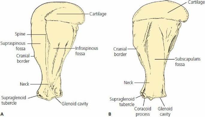
■ FIGURE 7-6 Scapula of the horse. A. Lateral view. B. Medial view. The supraspinatus and infraspinatus muscles occupy the respective fossas noted on the lateral surface. The suprascapular nerve that innervates these muscles is a branch of the brachial plexus and arises from the medial surface at the neck of the scapula. Nerve injury is a cause for atrophy (reduction in size and loss of function) of the associated muscles.
The pelvic girdle consists of the os coxae (hip bone), which unites ventrally with the opposite bone at the symphysis pelvis and articulates with the sacrum dorsally (Figure 7-7). The os coxae consists of the ilium, ischium, and pubis, which meet to form the acetabulum, the cavity that articulates with the head of the femur.
The ilium is the largest of the three components and projects craniodorsally from the acetabulum to articulate with the sacrum at the tuber sacrale. The ischium projects caudally and ventrally from the acetabulum and forms much of the caudal floor of the pelvic cavity. The pubis is the smallest of the three pelvic bones and forms the cranial part of the floor of the pelvic cavity. The obturator foramen is bounded by the pubis cranially and the ischium caudally. The tuber coxae and tuber ischiadicum vary in prominence among domestic animals, and in cattle are referred to as hook bones and pin bones, respectively. The obturator nerve passes through the obturator foramen to supply the adductor muscles of the pelvic limbs. Injury to the nerve during parturition (Figure 7-8) can cause failure to bring the pelvic limbs together. Also, because of the support given to the pelvic limbs by the pelvis (via the acetabulum), a fracture to the pelvis can result in an inability to stand.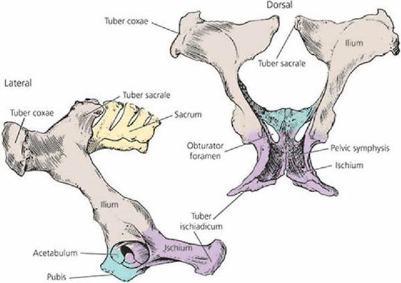
■ FIGURE 7-7 Pelvis of the ox. Lateral view (left) and dorsal view (right). (From Frandson RD, Wilke WL, Fails AD. Anatomy and Physiology of Farm Animals. 7th edn. Ames, IA: Wiley-Blackwell, 2009.)
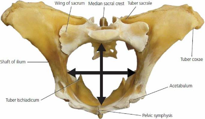
■ FIGURE 7-8 The pelvic bones of the cow (viewed from in front and somewhat below) through which the calf must pass at birth. The caudal aspect of the sacrum erroneously appears as an obstruction because of the view. The arrows indicate the greatest transverse and dorsoventral diameters of the pelvic girdle.
In general, among the domestic animals, the bones of the thoracic and pelvic limbs have like names but differ in size and shape and, for certain parts, differ in numbers. A comparison of the names for the bones between the thoracic and pelvic limbs is presented in Table 7-2. The relationship of these bones to each other is shown in Figure 7-9 and 7-10 for the thoracic limb.and pelvic limb, respectively, for comparison among the horse, ox, and pig.
Because of lameness problems, more attention is given to the anatomy of the limbs of horses, and the further description that follows will relate to that species. Whereas humans, cats, and dogs have five metacarpal bones (corresponding to the hand), horses have one major metacarpal (cannon bone), which is the third. The second and fourth exist as small bones alongside the third and are referred to as splint bones (Figure 7-11A). Fusion of these to the cannon bone along with excess bone formation may cause a lameness condition in horses that is known as splints. Distal to the cannon bone, the horse has a single digit (corresponds to the middle finger of humans) with three phalanges (Figure 7-11B). The names for the joints (articulations) between the cannon bone and the first phalanx (long pastern bone), between the first and second phalanx (short pastern bone), and between the second and third phalanx (coffin bone) are known as fetlock, pastern, and coffin joints, respectively. There are two proximal sesamoid bones and one distal sesamoid bone. Only the lateral proximal sesamoid is shown in Figure 7-11B. These sesamoid bones serve for the attachment of ligaments directed for more distal parts, and because of the articulation with adjoining bones, friction is reduced that would otherwise occur without their placement. The distal sesamoid is known as the navicular bone and is situated behind the junction of the second and third phalanges. Inflammation in the region of the coffin joint (location of the distal sesamoid bone) is called navicular disease. Because of the small size of the sesamoid bones, their name is derived from the small sesame seed. Not all sesamoid bones are small, however, and the patella (a sesamoid bone), which articulates with the femur, is an example.| TABLE 7-2 COMPARISON OF BONES OF THORACIC AND PELVIC LIMBS | |||
| THORACIC LIMB | PELVIC LIMB | ||
| PART OF LIMB | BONES | PART OF LIMB | BONES |
| Thoracic (shoulder) girdle | Scapula, clavicle, coracoid | Pelvic girdle | Sacrum pelvis: ilium, ischium, pubis |
| Brachium (arm) | Humerus | Thigh | Femur |
| Antebrachium (forearm) | Radius, ulna | Crus (true leg) | Tibia, fibula |
| Carpus (knee) | Carpal bones | Tarsus (hock) | Tarsal bones |
| Metacarpus (cannon and splint bones) | Metacarpal bones | Metatarsus (cannon and splint bones) | Metatarsal bones |
| Phalanges (digit) | Proximal, middle,.and distal phalanges Proximal and distal sesamoid bones | Phalanges (digit) | Proximal, middle, and distal phalanges Proximal and distal sesamoid bones |
From Frandson RD, Wilke WL, Fails AD. Anatomy and Physiology of Farm Animals. 7th edn. Ames, IA:.Wiley-Blackwell, 2009.
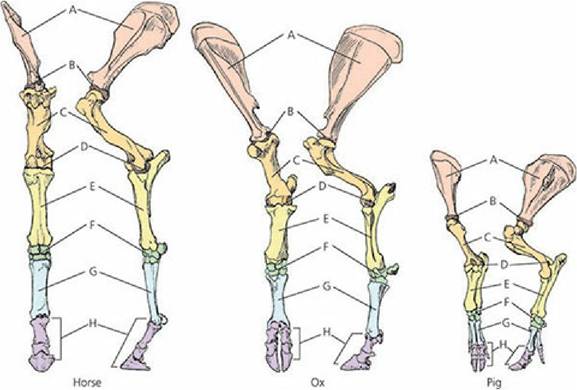
■ FIGURE 7-9 Comparison of anatomy of bones of the thoracic limb. A. Scapula. B.
Scapulohumeral (shoulder) joint. C. Humerus. D. Elbow joint. E. Antebrachium (radius and ulna). F. Carpus. G. Metacarpus. H. Digit (phalanges). (From Frandson RD, Wilke WL, Fails AD.
Anatomy and Physiology of Farm Animals. 7th edn. Ames, IA: Wiley-Blackwell, 2009.)
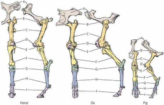
■ FIGURE 7-10 Comparison of anatomy of bones of the pelvic limb. A. Pelvis. B. Coxofemoral
(hip) joint. C. Femur. D. Patella. E. Stifle joint. F. Crus (tibia and fibula). G. Tarsus (hock). H. Metatarsus. I. Digit (phalanges). (From Frandson RD, Wilke WL, Fails AD. Anatomy and Physiology of Farm Animals. 7th edn. Ames, IA: Wiley- Blackwell, 2009.)
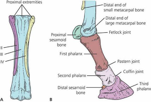
■ FIGURE 7-11 Metacarpal and phalangeal bones of the thoracic limb of the horse. A. Right metacarpal bones (palmar view). The third (III) or large metacarpal bone (cannon bone) is fully developed; the second (II) and fourth (IV) are much reduced and are commonly called the small metacarpal or splint bones. B. The phalanges and distal part of right metacarpal bones (lateral view).
The metatarsus and digits of the hindlimb are similar to the metacarpus and digits of the forelimb. The chief differences are in the form and size of the bones.
■