BONE STRUCTURE
1. What is another name for spongy bone?
2. Are the trabeculae (spicules) associated with compact or spongy bone?
3. How do trabeculae contribute to the strength of long bones?
4.
Differentiate between epiphysis, metaphysis, and diaphysis.5. How are bones associated with blood cell formation?
6. What is the epiphyseal plate? What parts of the bone does it separate?
7. What are periosteum and endosteum?
8. What percent of adult bone is water?
9. On a dry weight basis, what percent of adult bone is mineral content?
0. What part of bone is converted to gelatin when heated in aqueous solution?
11. What two elements are the major constituents of the mineral phase of bone?
2. What is the unit of structure of compact bone? Describe it.
3. What are lacunae and canaliculi? Where is bone interstitial fluid located?
4. What are interstitial and circumferential lamellae?
5. How are osteoprogenitor cells, osteoblasts, and osteocytes related?
6. Is the osteocyte more mature than the osteoblast?
7. How do osteocytes maintain communication with each other?
8. What is the name of the bone-resorbing cells? What is their origin?
The structure of a long bone (e.g., the femur) is shown in Figure 7-12. A longitudinal section is shown to reveal its inner structure. Compact and spongy characteristics are noted. Compact bone appears to be solid, whereas spongy bone (also called cancellous bone) has the appearance of a sponge. In spongy bone there are trabeculae (spicules) of mineralized tissue, and the empty spaces between the trabeculae occupy a considerable volume. In living animals, the regions between the trabeculae are filled with bone marrow. The rigidity and strength of long bones is attributable not only to the hardness of its compact bone but also to the scaffolding arrangement of the trabeculae, which are generally parallel to lines of maximum stress and, therefore, act as pillars for stress points (see Figure 7-12).
The epiphysis refers to either extremity of a long bone and the diaphysis is the cylindrical shaft situated between the two epiphyses. The metaphysis is the expanded or flared part of the bone at the ends.of the diaphysis. The diaphysis contains,the marrow (medullary) cavity that is surrounded by a thick-walled tube of compact bone. The medullary cavity or bone marrow is the site of blood cell production. A small amount of spongy bone may line the inner surface of the compact bone. The epiphyses consist chiefly of spongy bone with a thin outer shell of compact bone. The epiphyseal plate (also called physis) is composed of hyaline cartilage and represents the point of growth in a longitudinal direction. Hyaline cartilage is the ordinary type and is so named because its matrix (intercellular substance) is a glassy bluish-white (hyalos is the Greek word for glass) and is somewhat translucent. In mature bones, the cartilage has been replaced by bone and epiphyseal lines remain where the plate last existed. The contact area of the bone that articulates with its neighboring bone at a movable joint is covered with articular cartilage (described later in this chapter).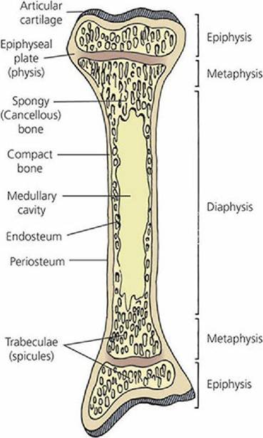
■ FIGURE 7-12 The structure of a long bone. The endosteum and periosteum designations refer only to their locations and do not reflect their cellular nature and extent. Note the parallel arrangement of the trabeculae to form scaffolding for maximum strength in response to its assumed load.
With the exception of the joint surfaces, all other outer surfaces of the bone are covered with periosteum. The periosteum is composed of an outer fibrous layer and an inner cell-rich layer containing osteoblasts (if bone formation is in progress) or other cells that can become osteoblasts in response to an appropriate stimulus (osteoprogenitor cells). Osteoblasts synthesize and secrete the organic substance of bone and participate in the mineralization of the organic matrix.
The periosteum is responsible for the increase in diameter of bones and also functions in the healing of fractures. The endosteum is the lining tissue of all surfaces of the bone that face the medullary cavity and also of the trabeculae of the spongy bone. It is only one cell thick, and the cells can become osteoblasts when stimulated.Composition of Bone
On a wet-weight basis, adult bone is approximately 25% water, 45% mineral, and 30% organic matter. Calcium constitutes about 37% of the mineral content and phosphorus about 18.5%. On a dry-weight basis, the mineral content is between 65% and 70%, whereas the organic fraction is 30% to 35%. The organic fraction is about 90% collagen, which is converted to gelatin when heated in aqueous solution. Several different elements are incorporated in the mineral phase of bone, but the major constituents are calcium and phosphorus.
Haversian Systems
Figure 7-13 is a three-dimensional illustration showing the appearance of both a crosssection and a longitudinal section of the shaft of a mature long bone. The channels that run parallel to the long axis of the bone are the Haversian canals, which contain blood vessels that communicate with blood vessels serving the external surfaces and marrow cavity. The latter blood vessels are perpendicular to the long axis of the bone and are contained within Volkmann canals. The unit of structure of compact bone is the Haversian system (also known as an osteon), which consists of a central Haversian canal surrounded by concentric layers of bone, the lamellae (Figure 7-14). Bone cells, the osteocytes, are contained within small cavities known as lacunae (little lakes). The osteocytes communicate with each other and with the Haversian canal through a branching network,of canals, the canaliculi. The interstitial,fluid for the osteocytes is contained within the lacunae and canaliculi. It diffuses through the canalicular network from the blood vessels in the canals for maintenance of the osteocytes.
Facilitation of fluid transport may be caused by periodic contraction of the osteocytes. Haversian systems are absent in spongy bone, but concentric lamellae with enclosed lacunae and osteocytes with intercommunicating canaliculi are present. In addition to the concentric lamellae that make up the Haversian system, other lamellar patterns occur in the form of interstitial lamellae and outer and inner circumferential lamellae (see Figure 7-13). The outer and inner circumferential lamellae are produced by the osteoblasts that cover the outer and inner surfaces of the bone while it is in the process of attaining its full width. During this time, Haversian systems develop, which gives the inner aspect of the outer circumferential lamellae and the outer aspect of the inner circumferential lamellae an interrupted appearance. Their uninterrupted aspects, however, give the outer and inner lamellar surfaces a smooth look. The interstitial lamellae are remnants of older Haversian systems or of circumferential lamellae.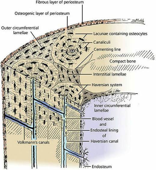
■ FIGURE 7-13 Three-dimensional diagram showing the appearance in cross section and longitudinal section of the components that enter into the structure of the cortex of the shaft of a long bone. (From Ham AW, Cormack DH. Histology. 8th edn. Philadelphia, PA: JB Lippincott Company, 1979.)
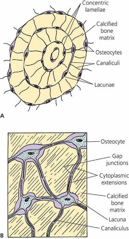
■ FIGURE 7-14 An osteon (Haversian system). A. Concentric lamellae, showing osteocytes within their lacunae and their communicating canaliculi. B. Cytoplasmic extensions of osteocytes into canaliculi for communication with other osteocytes.
Cells of Bone
Four different types of cells are associated with bone: osteoprogenitor cells, osteoblasts, osteocytes, and osteoclasts. The first of these, osteoprogenitor cells, comprise the population of cells in the innermost layer of the periosteum, the endosteal lining cells of the marrow cavities, and the lining cells of the Haversian canals and Volkmann canals.
Their stimulation leads to the more active secretory cell, the osteoblast. Where active bone formation is not occurring, the surfaces are covered by bone-lining cells, which are analogous to osteoprogenitor cells except that they represent a more quiescent state.The osteoblast is the differentiated bone-forming cell responsible for the production of bone matrix. Its secretion of collagen and ground substance makes up the initial unmineralized bone or osteoid. The osteoblast is also associated with calcification of the matrix.
The osteocyte is the mature bone cell and represents a transformed osteoblast. It is enclosed by the bone matrix that it had previously laid down as osteoid when it was an osteoblast. Osteocytes maintain the bone matrix and are able to synthesize and resorb matrix to a limited extent. They extend their cytoplasmic processes through the canaliculi to contact, by means of gap junctions, similar processes of neighboring cells. The gap junctions have a low electrical resistance that permit ionic and small molecule flow between cells. Communication among the osteocytes is thus possible such that the outermost cells, as well as those closest to blood vessels, can respond to stimuli (e.g., hormones). The osteocyte is smaller than in its previous state as an osteoblast because of reduced perinuclear cytoplasm. The appearance of osteocytes within their calcified bone matrix lacunae and their cytoplasmic extensions into the canaliculi is shown in Figure 7-14B.
Osteoclasts are large, motile, often multinucleated bone-resorbing cells. Their precursors are stem cells in blood-producing tissue of bone marrow and spleen. These stem cells differentiate into boneresorbing monocytes and then fuse with others to form the large multinucleated osteoclasts.
Osteoclasts are considered to be members of the diffuse mononuclear phagocytic system (MPS) (see Chapter 3).
Although osteoprogenitor cells, osteoblasts, and osteocytes are featured as distinct cell types, they should be regarded as different functional states of the same cell type.
■