GENERAL INTERNAL ANATOMY
The snake has evolved its body for crawling so has few external features. Elongation has also resulted in asymmetry of viscera with right-sided organs lying cranial to and being larger than left (Fig.
5.1). To explain the location of organs more easily it is best to divide the length of the snake roughly into three regions. The cranial region has the heart, trachea, esophagus, thyroid and proximal lung. The middle region has the stomach, liver, lung, spleen, and pancreas. The caudal region has the small and large intestines, kidneys and gonads.Cranial third
In all species of snake the heart lies cranioventral to the termination of the trachea, although it is mobile to allow for the passage of large food items. The thyroid gland is just cranial to the heart. The thymus gland (which does not involute in adults) is thin and lies on the trachea proximal to this. The rostral parathyroids lie near the angle of the jaw while the caudal pair lies near the thymus, just rostral to the heart (McCracken 1999) (Figs. 5.2 and 5.3).
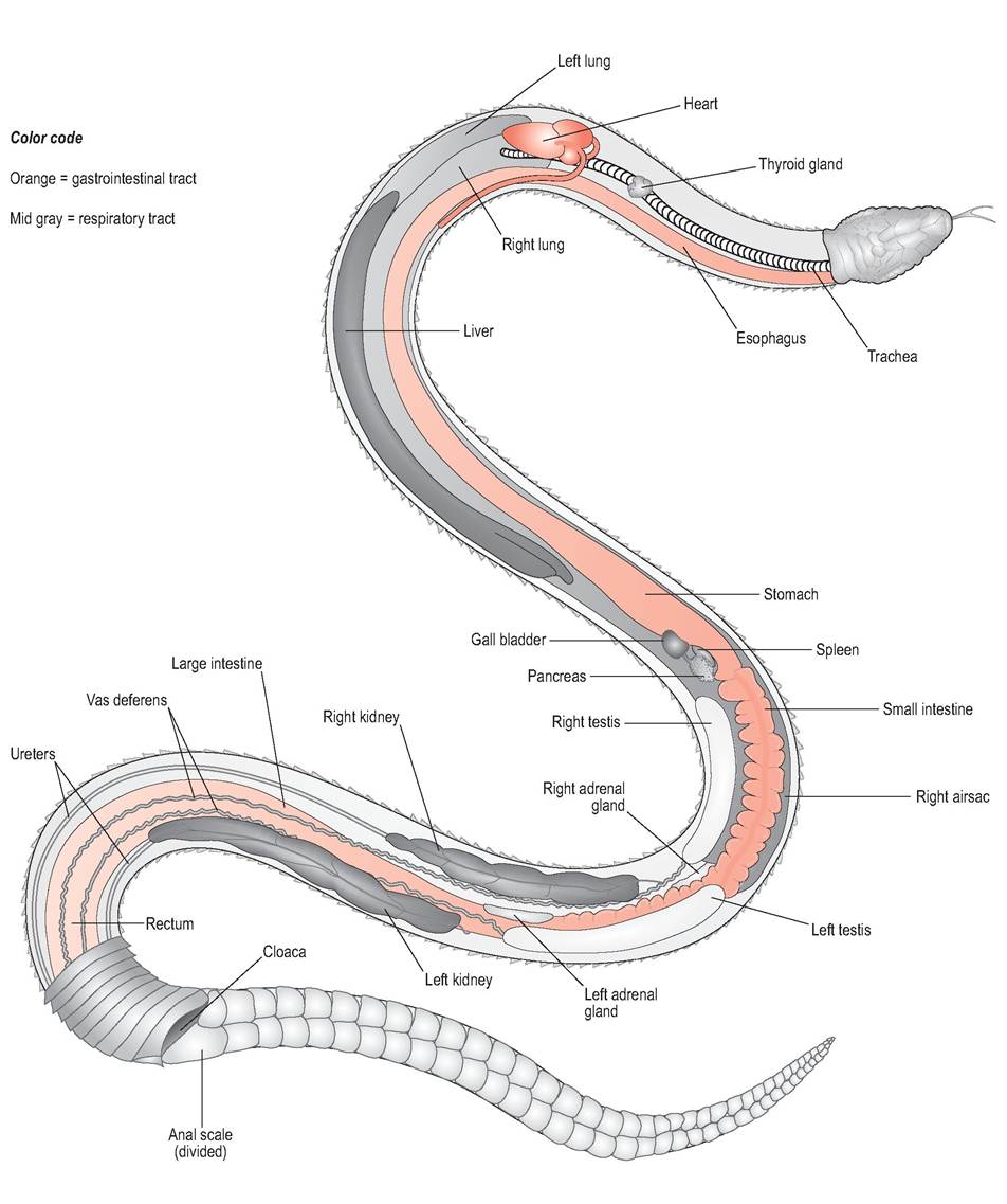
Figure 5.1 • Internal anatomy of the snake (male) - the celomic fat pads have been removed to show caudal viscera. Note the location of the gall bladder some distance from the liver.
Colour code
Orange = gastrointestinal tract
Mid gray = respiratory tract
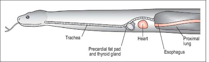
Figure 5.2 • Internal anatomy of cranial third showing location of trachea, thyroid, heart, proximal lung (right) and esophagus.
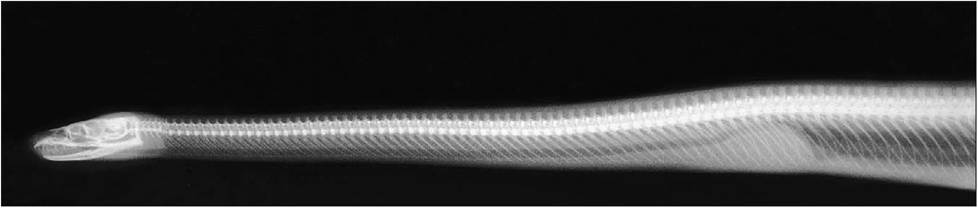
Figure 5.3 • Lateral radiograph of cranial third of the snake.
Middle third
Caudal third
The gall bladder lies near the pylorus of the stomach and is located some distance from the posterior pole of the liver.
In some species the spleen and pancreas are fused into a splenopancreas and lie adjacent to the gall bladder, forming an organ triad. The right ovary lies either near to this triad or just caudal to it (McCracken 1999) (Figs. 5.4 and 5.5). The right and left gonads occur in sequence, followed by the right and left kidneys. The pink adrenal glands can be visualized medial to the respective gonads and lie in the mesorchium/mesovarium. The intestines are linear and in Boidae a cecum is visible at the junction of the small intestine and colon. Celomic fat bodies lie ventral to all the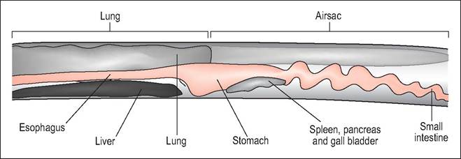
Figure 5.4 • Internal anatomy of middle third of the snake showing liver, lung, stomach, spleen, and pancreas.
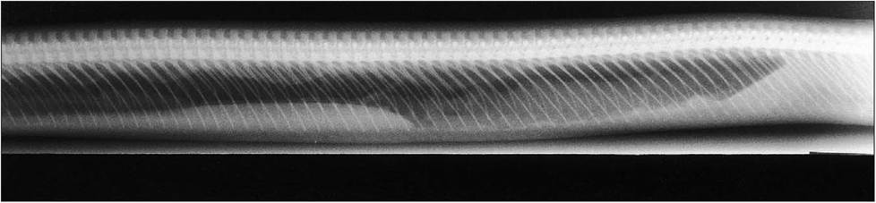
Figure 5.5 • Radiograph of middle third of the snake.
viscera, starting at the level of the gall bladder and extending caudally to the level of the cloaca (McCracken 1999) (Figs. 5.6 and 5.7).