SKELETAL SYSTEM
Skull
The snake has the most kinetic skull of all, with extremely flexible and mobile bones in all parts. It has no temporal arch, interorbital septum or middle ear cavity. Instead, its intricate design enables a reptile with a small mouth to eat enough large prey to sustain its length.
The skull is modified so that all the tooth-bearing bones of the skull are able to move independently; the braincase is heavily ossified to protect it from protesting prey (Liem et al. 2001a) (Fig. 5.8).Snakes have no mandibular symphysis; instead, flexible skin allows the jaw bones to move apart and forward or backward (Figs. 5.9 and 5.10). The quadrate bone which articulates with the lower jaw and palatomaxillary arch also has a very loose articulation. This becomes rigid when under tension but extremely flexible when relaxed (Fig. 5.10). More advanced viperid snakes have elongate quadrate bones slanting backwards and outwards giving them the notorious triangular shaped head. Many also have an articulation between the prefrontal and maxillary bones (Bellairs 1969b; Pough 1998a; Pough et al. 2002) (Fig. 5.11).
Each half of the skull works separately and this independence allows the snake to literally walk its jaw along large prey (Fig. 5.12). The left half of the upper and lower jaw can be moved and then clamped allowing the right side to advance forwards. Snakes often yawn after a meal to allow their jawbones to reposition themselves.
Vertebrae
There are often up to 400 vertebrae precloacally, each with its own pair of ribs and large axial skeletal muscles. Each vertebra has five separate articulations with its fellow vertebrae and this, combined with the large number of vertebrae, results in a very flexible backbone (Hoffstetter & Gasc 1970). The hypaxial and epaxial muscles extend along these vertebrae by an interlocking system of muscle chains and tendons, thus adding to the snake's flexibility.
The intercostal and hypaxial muscles not only help in locomotion but also in the passage of prey for digestion and in respiration.Snakes have no distinct cervical region but the first two cervical vertebrae lack ribs. There is no sternum or costal cartilages so each rib pair attaches by muscles to the inner surface of the ventral scales. Post cloaca there are no distinct ribs but vertebral processes fork ventrally and dorsally to protect the lymph hearts (Figs. 5.13 and 5.16). The tail is always shorter than the trunk (Hoffstetter & Gasc 1970).
GENERAL INTEREST
The cobra has long curved ribs on its cervical vertebrae, which can be rotated outwards causing a fold of loose skin to spread. This hood is then inflated with air from the lungs.
Locomotion
Snake locomotion involves two kinetic types: undulating and sidewinding (Fig. 5.14). They often combine several methods of undulation according to the local terrain (Bellairs 1969a).
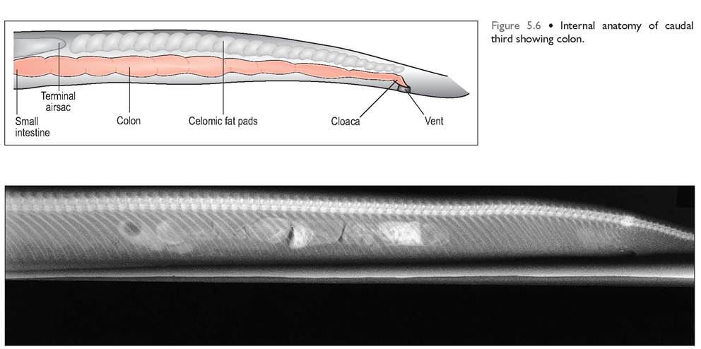
Figure 5.7 • Radiograph of caudal third of the snake. Other viscera are not visible radiographically as they merge with the large celomic fat pads in this area. Note the absence of ribs post cloaca.
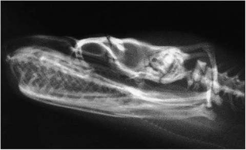
Figure 5.8 • Lateral radiograph of skull of common boa (Boa constrictor) showing flexible serpentine skull. Note heavily ossified braincase, backwardly pointing teeth and mobile quadrate bone.
Lateral undulation
This is when the snake wriggles laterally. When the body comes in contact with an object or rough surface it thrusts itself forward. As they need a minimum of three contact points, snakes would have difficulty advancing on very smooth surfaces like glass. Long, thin snakes like racers can move faster because they can make more curves and more thrust. During swimming snakes use this movement, pushing against the resistance of the water (Pough et al.
2002).Rectilinear
Rectilinear locomotion is where a snake moves forward in a straight line on its ribs, causing a wave effect. This method of motion is used by thick-bodied snakes like boas and pythons and is less conspicuous than other forms. It is also used for stalking prey. The ventral scales are loosely attached and linked to the ribs by segmental muscles. Contraction of these muscles helps to draw the snake forward (Pough et al. 2002).
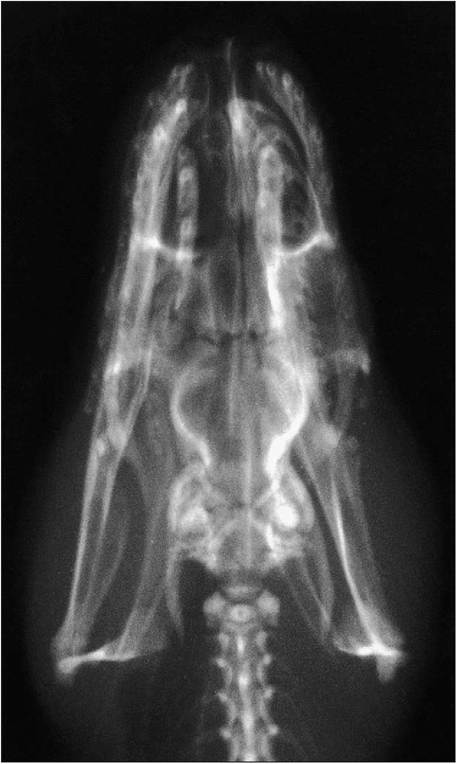
Figure 5.9 • Dorsoventral radiographs of above demonstrating lack of mandibular symphysis.
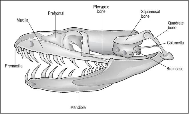
Figure 5.10
Skull of simple snake.
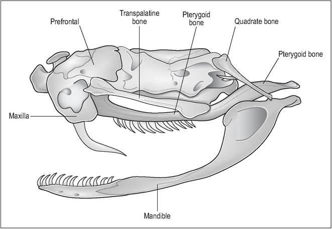
Figure 5.ll • Advanced snake skull as seen in vipers demonstrating the shortened maxilla. The prefrontal bone can be raised like a hinge to rotate the front fangs for striking.
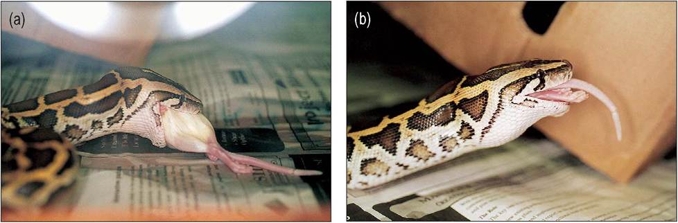
Figure 5.12(a&b) • Burmese python (Python molurus) eating a rat. The snake’s skull is modified to allow an animal with a small head to consume prey large enough to sustain their length. The tooth-bearing bones of the skull are able to move independently of each other, allowing the snake to “walk” its jaw over large prey.
Concertina
The concertina method is used mainly by burrowing snakes. This is where the snake bunches a few s-shaped loops against the body wall and then straightens out at the front to move forward (Pough et al. 2002).
Sidewinding
Sidewinding is a specialized locomotion devised by snakes such as rattlesnakes and vipers that live in deserts. It involves throwing the head forward while at the same time throwing a loop of body forward, leaving a characteristic ‘J’- shape behind. The body actually moves at right angles to the direction the snake is travelling and is the fastest method of maneuvering through loose sand (Pough et al. 2002).
Spurs
With elongation, the snake has lost its pectoral girdle to facilitate the swallowing of large prey. Some of the more primitive Boidae have retained pelvic vestiges, “spurs”, and these can be seen on either side of the vent (Figs. 5.15 and 5.16). These short bones are covered in keratin and articulate with a longer bone lying within the rib cage. An attached muscle serves to flex and extend the spurs, which are used during courtship and mating (Evans 1986).