GROSS ANATOMY OF THE KIDNEYS AND URINARY BLADDER
1. Study the shape of kidneys of different species.
2. What is the location of the kidney cortex and medulla? What is the renal hilus and renal pelvis?
3. What is the difference between the ureter and the urethra?
4.
What is the relationship between the ureterovesicular junction and prevention of backflow of urine from the bladder to the kidney?5. Describe the innervation to the kidneys.
The kidneys are paired organs suspended from the dorsal abdominal wall by a peritoneal fold and the blood vessels that serve them. They are located slightly cranial to the midlumbar region (Figure 11-1). Because they are separated from the abdominal cavity by their envelopment of peritoneum, they are called retroperitoneal structures. Blood is carried to each kidney by a renal artery and venous blood is conveyed away from each kidney by a renal vein. The renal artery arises directly from the aorta and the renal vein empties directly into the caudal vena cava (Figure 11-2).
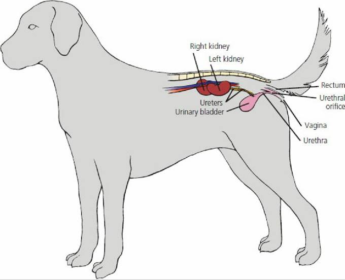
■ FIGURE 11-1 Side view of female dog showing general location of kidneys, ureters, urinary bladder, urethra, urethral orifice, and vagina.
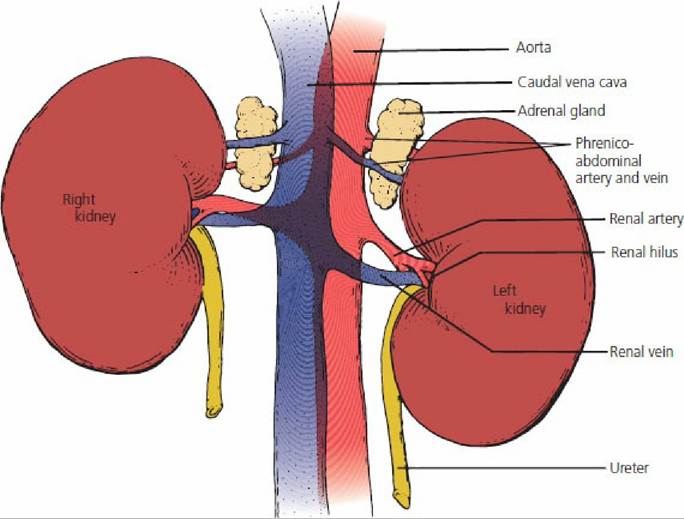
■ FIGURE 11-2 Ventral view of canine kidneys showing renal arteries, veins, and ureters and their positions relative to the aorta, vena cava, and adrenal glands.
The kidney is described as a bean-shaped structure for most domestic animals. In the horse, however, it is described as heart-shaped and in cattle it is lobulated (Figure 11-3). If a midsagittal cut is made through the kidney (Figure 11-4), an outer cortex and an inner medulla are visible. The striations of.the medulla are formed by the anatomic arrangement of the major parts that occupy the medulla, the loop of Henle of long-looped nephrons and the medullary portion of the collecting tubules (see later section on the nephron).
The medullary portions of the collecting tubules are known as collecting ducts. The renal hilus is the indented area on the concave edge of the kidney through which the ureter, blood vessels, nerves, and lymphatics enter or leave. The renal pelvis (see Figure 11-4) is the expanded origin of the ureter within the kidney. The final discharge of urine from the many collecting ducts is received by the renal pelvis. Innervation to the kidney is provided by the sympathetic (adrenergic) division of the autonomic nervous system. The postganglionic renal nerves enter the hilus of the kidney in association with the renal artery and vein and provide adrenergic innervation to the renal vasculature, all segments of the nephron, and the juxtaglomerular (JG) granular cells. The ureter is a muscular (smooth muscle) tube that conveys urine from the renal pelvis to the urinary bladder. The ureter enters the bladder at an oblique angle (ureterovesicular junction), thus forming a functional valve to prevent backflow when the bladder is filling (Figure 115). The urinary bladder is a hollow, muscular (smooth muscle) organ that varies in size depending on the amount of urine it contains at any one time. The smooth muscle of the urinary bladder is known as the detrusor muscle. The epithelial cell lining of the bladder accommodates for the change in size and is known as transitional epithelium (see Chapter 1). When the bladder is empty, the cells appear to be piled on one another, giving it a stratified (layered) appearance. A transition occurs on filling so that the piled-up appearance gives way to a thinner epithelial stratification.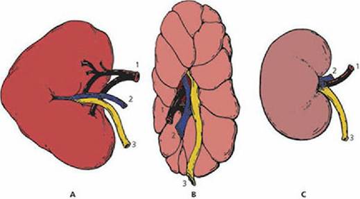
■ FIGURE 11-3 Right kidney from various species. A. Horse. B. Cattle. C. Sheep. These represent heart-shaped, lobulated, and bean-shaped kidneys, respectively. (1) Renal artery; (2) renal vein; (3) ureter.
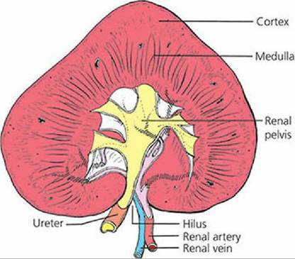
■ FIGURE 11-4 Midsagittal plane of horse kidney showing cortex, medulla, renal pelvis, hilus, ureter, renal artery, and renal vein.
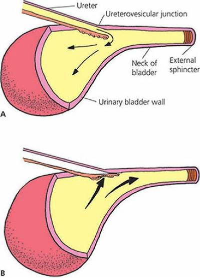
■ FIGURE 11-5 Ureterovesicular junction (oblique entrance of ureter into the urinary bladder). A. Urine is conveyed to the urinary bladder from the renal pelvis by peristalsis and enters at the ureterovesicular junction. B). During micturition (emptying of the urinary bladder), urine is directed through the neck of the bladder to the urethra. Urine does not reenter the ureter because the ureterovesicular junction is closed by the hydrostatic pressure of urine associated with contraction of the detrusor muscle of the bladder wall.
The neck of the bladder is the caudal continuation of the bladder leading to the urethra. The smooth muscle in the neck is mixed with a considerable amount of elastic tissue and functions as an internal sphincter.
The urethra is the caudal continuation of the neck of the bladder. It conveys the urine from the bladder to the exterior (Figure 11-6). The external sphincter lies beyond the neck; it is composed of skeletal muscle that encircles the urethra at this point. The functional boundary between the bladder and the urethra is represented by this sphincter.
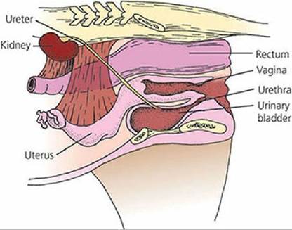
■ FIGURE 11-6 Midsagittal plane of the mare pelvis showing positions of the urinary bladder and urethra relative to other organs.
Prevention of urine escape while the bladder is filling is provided for by contraction of the external sphincter and by tension passively exerted by the elastic elements in the neck of the bladder. When urine is expelled from the bladder, the external sphincter relaxes and the bladder muscles contract. The bladder muscle contraction opens its neck into a funnel shape. The contraction not only forces urine into the urethra but, because of the muscle fiber arrangement, widens the beginning of the urethra.