THE NEPHRON
1. Do large-breed dogs have significantly greater numbers of nephrons in their kidneys than small-breed dogs?
2. What is the difference between a cortical nephron and a Juxtamedullary nephron?
3.
What are the components of the nephron (in order) from the glomerulus to the inner medullary-collecting duct?4. What are the components of the JG apparatus?
The functional unit of the kidney is the nephron. An understanding of nephron function is essential for understanding kidney function. Nephron numbers vary considerably among species, and approximate numbers for several species are given in Table 11-1. Within a species the nephron numbers are relatively constant. Considering the differences in size among various breeds of dogs, it might be thought that the kidneys of large-breed dogs would contain more nephrons than the kidneys of small-breed dogs. This is not the case, however, and the larger kidney size in large dogs is compensated for by their having larger nephrons rather than more nephrons.
| TABLE 11-1 APPROXIMATE NUMBER OF NEPHRONS.IN EACH KIDNEY FOR. SEVERAL DOMESTIC ANIMALS AND HUMANS | |
| SPECIES | NEPHRONS/KIDNEY |
| Cattle | 4,000,000 |
| Pig | 1,250,000 |
| Dog | 415,000 |
| Cat | 190,000 |
| Human | 1,000,000 |
The mammalian kidney has two principal types of nephrons, identified by: (1) the location of their glomeruli and (2) the depth of penetration of the loops of Henle into,the medulla. Those nephrons with glomeruli in the outer and middle cortices are called cortical or corticomedullary nephrons.
They are associated with a loop of Henle that extends to the junction of the cortex and medulla or into the outer zone of the medulla. Those nephrons with glomeruli in the cortex close to the medulla are known as juxtamedullary nephrons. Juxtamedullary nephrons are associated with loops of Henle that extend more deeply into the medulla; some extend as deep as the renal pelvis. The relationship of each nephron type to the cortex and medulla is shown in Figures 11-7 and 11-8. The juxtamedullary nephrons are those that develop and maintain the osmotic gradient from low to high in the outer medulla to the inner medulla, respectively. The percentage of nephrons having long loops of Henle (juxtamedullary nephrons) varies among animal species and ranges from 3% in the pig to 100% in the cat. In humans the percentage of long-looped nephrons is about 14%. The tubular fluid from both nephron types enters the collecting tubules and collecting ducts, where it is exposed to the effects of medullary osmotic gradients as it proceeds to the renal pelvis.
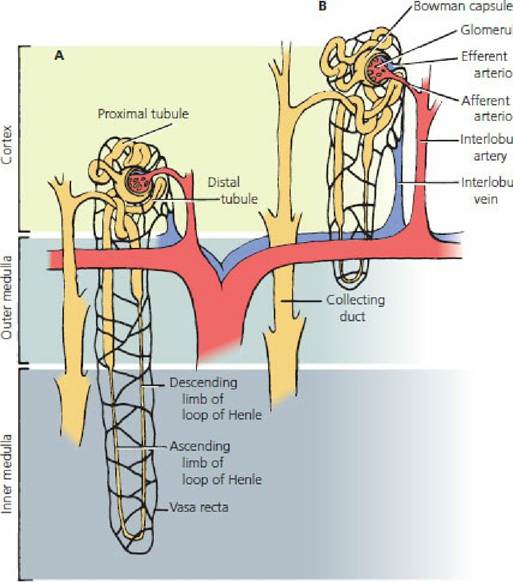
■ FIGURE 11-7 Types of mammalian nephrons. A. Juxtamedullary (long-looped) nephron. B. Cortical nephron.
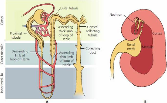
■ FIGURE 11-8 A. Component parts of a Juxtamedullary nephron (mammalian) relative to their locations in the cortex and medulla. B. Midsagittal section of the kidney showing the location of a Juxtamedullary nephron (exaggerated size) relative to the cortex, medulla, and renal pelvis.
Nephron Components
A typical nephron and its component parts are shown in Figure 11-Q. The glomerulus is the tuft of capillaries through which filtration is accomplished. The afferent arteriole conducts blood to the glomerulus and the efferent arteriole conducts blood away from the glomerulus. Blood leaving through the efferent arterioles is redistributed into another bed of capillaries known as the peritubular capillaries; these perfuse the nephron tubules.
The vasa recta are capillary branches from the peritubular capillaries associated with the long-looped nephrons. After perfusion of the kidneys, blood is returned to the caudal vena cava by the renal veins.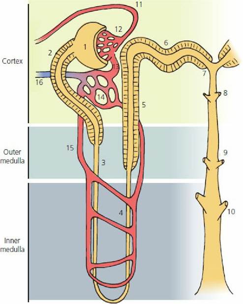
■ FIGURE 11-9 The functional nephron with blood supply. A Juxtamedullary nephron is shown so as to display the vasa recta. (1) Bowman’s capsule; (2) proximal tubule; (3) descending limb of loop of Henle; (4) thin ascending limb of the loop of Henle; (5) thick ascending limb of the loop of Henle; (6) distal tubule; (7) connecting tubule; (8) cortical collecting tubule; (9) outer medullary collecting duct; (10) inner medullary collecting duct; (11) afferent arteriole; (12) glomerulus; (13) efferent arteriole; (14) peritubular capillaries; (15) vasa recta; (16) to renal vein. The thick ascending limb of the loop of Henle becomes a distal tubule when it passes between the afferent and efferent arteriols at the glomerulus (location of the macula densa).
Nephron Tubules and Ducts
Filtrate from the glomerulus is collected by Bowman’s capsule and is subsequently directed through the proximal tubule, loop of Henle, and distal tubule. The distal tubule empties into a cortical collecting tubule. A cortical collecting tubule is not unique to a single nephron because it receives tubular fluid from the convoluted portion of several distal tubules. When the collecting tubule turns away from the cortex and passes down into the medulla, it is known as a collecting duct. Successive generations of collecting ducts coalesce to form progressively larger collecting ducts. The tubular fluid is finally discharged from,the larger collecting ducts into the pelvis,of the kidney, and is conveyed from there by the ureters to the urinary bladder for storage until discharge through the urethra. A summary of nephron component parts encountered by the glomerular filtrate as it becomes tubular fluid and finally urine with final discharge through the urethra is shown in Figure 11-10.

■ FIGURE 11-10 Summary of kidney blood flow and tubular fluid flow as it applies to the nephron. After removal of the filtration fraction of plasma at the glomerulus, the remaining blood that enters the efferent arteriole is distributed via peritubular capillaries to the nephron, as shown. The fraction of plasma filtered at the glomerulus enters Bowman’s capsule as a glomerular filtrate. It continues through the nephron tubules and ducts as tubular fluid. The tubular fluid is subjected to reabsorption and secretion and enters the renal pelvis as urine. Urine is finally evacuated from the urinary bladder by micturition.
Loop of Henle
The loop of Henle is composed of three segments: the thin descending limb, the thin ascending limb, and the thick ascending limb. Their relative thicknesses are a result of differences in the epithelial cells and do not refer to changes in lumen diameter. The thin segment for each loop is continuous with the thin segment of the other at the hairpin curve. The descending limbs of cortical nephrons only go as deep as the outer aspect of the outer medulla. The juxtamedullary nephrons have descending limbs of loops of Henle that can extend to the renal pelvis. The thin segment of the descending limb is a straight tubule continuous from the proximal tubule and is followed after its hairpin turn by the thin ascending limb. The thick segment of the ascending limb is a straight tubule continuous from the thin ascending limb. The thick segment of the ascending limb of the loop of Henle returns in its ascent to its glomerulus of origin, passes between the afferent and efferent arteriole, and proceeds from there as the distal tubule to its cortical collecting tubule.
Juxtaglomerular Apparatus
The junction of the distal tubule and glomerulus is known as the juxtaglomerular (JG) apparatus (Figure 11-11). There are characteristic cell types at this location.
In the tubule, the cells are collectively known as the macula densa; in the afferent and efferent arterioles, they are called the JG cells and the cells located between the macula densa and the arterioles are known as extraglomerular mesangial (lacis) cells. The JG apparatus is associated with regulating the amount of blood flowing to the kidney, the amount of filtration, and,the secretion of renin, an enzyme involved in the formation of the hormone angiotensin II (a vasoconstrictor).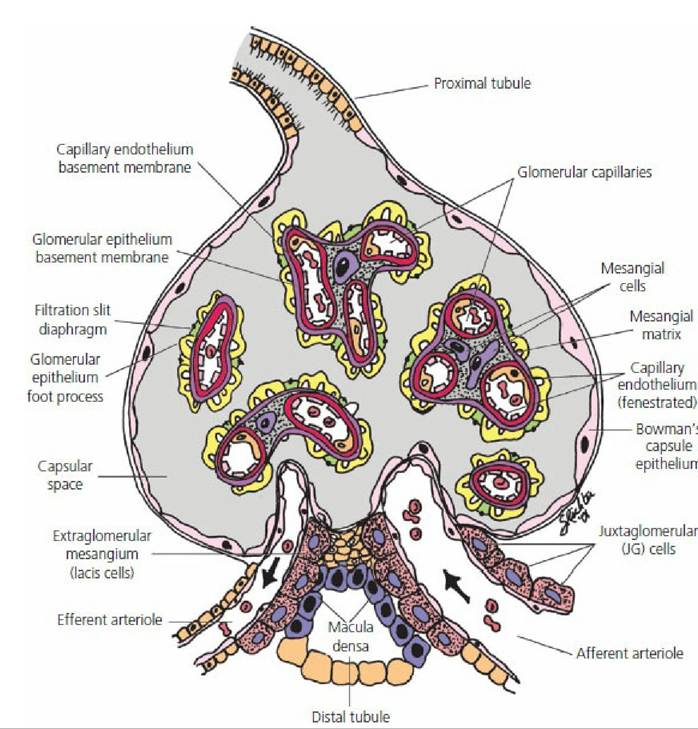
■ FIGURE 11-11 The juxtaglomerular (JG) apparatus. The JG apparatus is located at the junction of the distal tubule and its glomerulus of origin. It is associated with regulation of blood flow and filtration fraction for the nephron and with the secretion of renin, an enzyme involved in the formation of angiotensin II. Structures within the capsular space (Bowman’s capsule) appear as independent structures because of the transverse section view. Structurally, they are continuous with each other and with the afferent and efferent arterioles. (From Reece WO. Kidney function in mammals. In: Reece WO, ed. Dukes’ Physiology of Domestic Animals. 13th edn. Ames, IA: Wiley- Blackwell, 2015.)
■