HEART SOUNDS
1. What are the heart sounds and with what are they associated?
Listening to the heart (cardiac auscultation) enables the listener to hear the sounds that accompany contraction of heart muscle and the sounds associated with closure of the heart valves.
These are repeated for each cardiac cycle. The more pronounced sounds are those associated with valve closings, but contracting muscle also makes a sound.The first heart sound resembles the word “lub” and the second heart sound resembles “dub.” They usually occur one after the,other - lub-dub, lub-dub, lub-dub, and so on. The first sound is produced when the ventricles contract and the A-V valves close. The second sound is produced when ventricular relaxation begins and the semilunar valves close. The abrupt closing of the valves and their potential for making sounds can be visualized. A third heart sound can sometimes be detected on a phonocardiogram (recording of heart sounds); this occurs toward the end of rapid filling of the ventricles. Heart sounds are useful as diagnostic aids because the heart valves can become diseased and might not close completely. When this happens, blood leaks through the valves and the turbulence of the leakage is heard as some variation of a “shhh” sound after the lub or dub. Abnormal heart sounds are called heart murmurs and usually result from valve disorders. The simultaneous recording of a phonocardiogram, electrocardiogram, respiration, and blood pressure is illustrated in Figure 9-26 and provides a review of the relationship of the ECG waves with ventricular systole and associated pressures and of the resulting valve closings with their associated sounds.
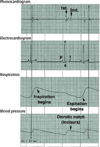
■ FIGURE 9-26 Simultaneous recording of electrocardiogram (lead II), phonocardiogram, respiration, and blood pressure of a dog. The correlation of events is represented by correlation line A (first heart sound, lub; ECG; blood pressure) and B (second heart sound, dub; blood pressure). For line A: immediately after depolarization of the ventricles, heart contraction begins, the first heart sound is perceptible, and blood pressure begins to increase. For line B: ventricular relaxation begins, blood pressure decrease begins, and semilunar valves close. Valve closure produces the second heart sound and causes a momentary blood pressure bounce upward (dicrotic notch). Respiratory sinus arrhythmia (an example of the Bainbridge reflex) is shown by correlating the inspiratory phase of the breathing cycle (blood flow to right atrium increases) with increased heart activity (R-R interval decreased). Paper speed, 25 mm/s; 1 cm = 1 mV. See Figure Q-25 for a study of the grid measurements.
■