Hormones of Pregnancy
Progesterone
Progesterone has several actions that are essential for maintaining a normal pregnancy. These include (1) providing negative feedback to the hypothalamus to inhibit any further estrous cycles, (2) inhibiting the smooth muscle of the uterus to permit the attachment and development of the fetus, and (3) assisting with maintenance of the contractility of the cervix to protect the uterine environment.
Plasma levels and sources of progesterone differ among species and among stages of pregnancy. in all domestic animals, the initial source
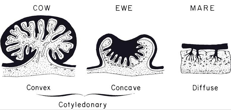
Figure 28-5. Placental attachments of cow, ewe, and mare. Villi from chorioallantois (black) invaginates into crypts in maternal uterine epithelium (stippled) in caruncles in the cow and the ewe and in diffuse locations in the mare.
of the necessary progesterone is the corpus luteum, and in some species (e.g., cow and sow), the corpus luteum remains the primary source throughout pregnancy. in other species (e.g., mare and ewe), the initial corpus luteum can be removed after secondary sources produce enough progesterone to maintain pregnancy. These sources include secondary or accessory corpora lutea and the placenta in the mare; the placenta is the secondary source in the ewe.
Equine Chorionic Gonadotrophin
The mare appears to be unique among domestic species in that the equine placenta is the source of a protein hormone that acts similarly to the luteinizing hormone, the source of which is the pituitary. Secretion of equine chorionic gonadotrophin (eCG, formerly known as pregnant mare serum gonadotrophin, or PMSG) begins after about a month of gestation and continues until about 4 months of gestation. During this period, follicular development occurs on the ovary of the pregnant mare, and ecG promotes the luteinization of these follicles. These accessory corpora lutea provide secondary sources of progesterone.
Trophoblastic cells of fetal origin found in specialized structures termed endometrial cups are the source of the ecG. These structures are seen as small, raised circular areas with a central depression on the endometrial surface of the pregnant uterus. The trophoblastic cells are found in association with the endometrium in these areas.
Relaxin
Relaxin is a protein hormone secreted by the corpus luteum in some species (sow and cow) and the placenta in others (bitch and mare). The primary function of relaxin is preparation for parturition and ultimately lactation. Relaxin contributes to the opening of the cervix and the relaxation of the muscles and ligaments associated with the birth canal to facilitate the passage of the fetus. in some species, peak secretion of relaxin occurs just prior to parturition. Gradual increases in relaxin during the latter part of gestation also facilitate mammary gland development to prepare for lactation.