Hypoxemia in an Anesthetized Clydesdale Horse
History. A 2-year-old, 750-kg Clydesdale horse is presented for removal of a testicle that has been retained in the abdomen, a procedure that requires anesthesia. You know that anesthesia of heavy draft horses can lead to gas exchange problems, and therefore you have an anesthesia machine available to provide ventilation and to supplement the horse with extra oxygen.
The horse is anesthetized with a short-acting intravenous anesthetic; an endotracheal tube is inserted; and the horse is connected to the anesthesia machine and allowed to breathe oxygen containing isoflurane for anesthesia. Ventilation is not assisted.Thirtv minutes after the induction of anesthesia, the veterinary technician takes an arterial blood sample to monitor the horses gas exchange. PaO2 is 70 mm Hg, and Paco2 is 65 mm Hg. Are you satisfied with the results of the blood gas analysis? If not, what can be done to improve gas exchange?
Comment. The elevation of Paco2 from the normal value of 40 mm Hg to 65 mm Hg shows that the horse is suffering from alveolar hypoventilation; that is, the ventilation received by the alveoli is insufficient to remove the carbon dioxide being produced by the horse. This is probably a result of depression of the central nervous system by the anesthetic gases, so that the drive to breathe is reduced. In addition, the positioning of the horse on its back for removal of the retained testicle causes the heavy viscera to push on the diaphragm, making it difficult for the horse to ventilate. Alveolar hypoventilation in an anesthetized animal can be corrected by the use of positivepressure ventilation. You have a ventilator as part of the anesthesia machine and choose to ventilate the horse to return the Paco2 to acceptable levels.
The Pao2 of 70 mm Hg shows that the horse has considerable problems in exchanging oxygen.
Although a Pao2 of 70 mm Hg is sufficient almost to saturate hemoglobin and would not be considered particularly low in an animal breathing air, it is very low in an animal breathing 100% oxygen. When animals breathe oxygen, PAo2 is greater than 600 mm Hg: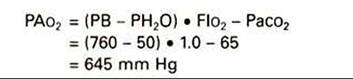
If the lung is functioning ideally, arterial oxygen should also be close to 600 mm Hg. In this horse, Pao2 is only 75 mm Hg, so AaDo2 is 645 - 75, that is 570 mm Hg.
This huge AaDo2 is not unusual in large, anesthetized mammals. The positioning of the horse on its back with the consequent weight of the viscera pushing forward on the diaphragm and compressing the lungs can lead to severe V/Q inequalities. Parts of the dependent lung are unable to ventilate, although they continue to receive blood flow and therefore become right-to-left shunts. These right-to-left shunts result in severe arterial hypoxemia. As long as the Pao2 is sufficient to saturate hemoglobin, the horse is in no danger. The dangerous point is during recovery from anesthesia. The horse must be supplemented with oxygen until it is sufficiently conscious to be able to rest on its sternum unaided and eventually to stand. Returning to these postures eliminates right-to-left shunts, restores the V/Qdistribution to normal, and improves gas exchange.