IMMUNE SYSTEM
The lymphatic circulation is intimately associated with the circulatory system in chelonia and can pose a major complication in blood sampling. The deep jugular trunk passes close to the jugular vein and a subpubic sinus, which drains the tail, cloaca, and caudal limbs can affect blood samples from the dorsal coccygeal vein.
The skin has a network of lymphatic vessels that are widely meshed, becoming superficial near the attachment of skin to the shell. The orbits have two lymphatic sinuses that extend into both eyelids. A single pair of lymphatic hearts lies at the most caudal part of the trunk, deep to the last vertebral shield of the carapace.
Some species, like the Red-eared sliders (Trachemys scripta) and European pond turtle (Emys orbicularis), have
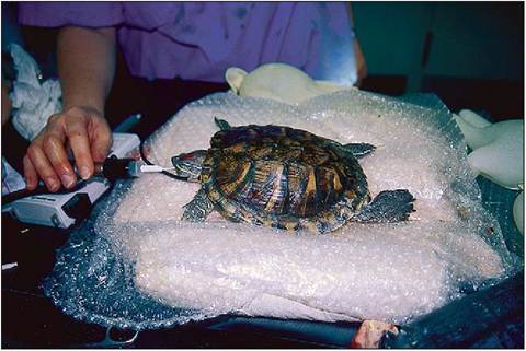
Figure 3.19 • Induction of anesthesia can be difficult in aquatic species.
Upper respiratory tract
Chelonia breathe with their mouth closed. Air enters via the external nares into the nasal cavity and passes through the partial hard palate to the pharynx. The glottis is easily visible at the back of the short, fleshy tongue. The trachea has complete cartilaginous rings. In Cryptodira species the trachea is very short and bifurcates rapidly to allow for head retraction.
Lower respiratory tract
The lungs are spongy and occupy a large volume in the dorsal half of the body cavity, although their volume is reduced to one fifth when the head and limbs are retracted (Gans & Hughes 1967). They are attached dorsally to the periosteum of the carapace and tightly against the pectoral and pelvic limb girdles. They are not surrounded by a pleural cavity and are only separated from the ventral cavity and viscera by a thin non-muscular postpulmonary septum, which plays no active part in respiration (Murray 1996b; Perry 1989).
Internally, chelonian lungs are surprisingly advanced for such a primitive reptile (Fig. 3.20). They are multicameral,
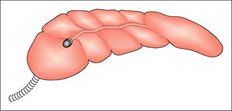
Figure 3.20 • Schematic drawing of multicameral lungs of Chelonia. Despite their ancient lineage and primitive appearance chelonians have more advanced lungs than snakes and some lizards.
as in the Monitor lizard, with a single intrapulmonary bronchus radiating into a network of bronchioles and highly vascular faveoli (Perry 1989). However, unlike in the Monitor lizard, the lungs are confined only to the dorsal half of the body, with the heart lying cranially near the pectoral inlet (Figs. 3.21-3.23) (see Chapter 4).
CLINICAL NOTE
Remember, most chelonians have a very short trachea, so when intubating do not insert the endotracheal tube too far as you may be intubating one primary bronchus instead (Murray 1996a; Gans & Hughes 1967).
Ventilation
The absence of a diaphragm and the fact that they have modified their ribs, sternum, and vertebrae into a hard shell means there is no expandable chest (McCutcheon). Therefore, in order to breathe, chelonians have developed strong trunk muscles, which expand and contract the lungs with active inspiration and expiration (Fig. 3.24) (Gans & Hughes 1967; McCutcheon 1943; Pough 1998a). It is the action of these antagonistic muscles moving the ventral postpul- monary septum that draws air in and out of the lungs. Terrestrial species breathe regularly but aquatic species can only breathe when they surface for air, otherwise the high volume of air would act as a natural buoyancy aid.
CLINICAL NOTE
When Chelonia have their head retracted inside their shell they can no longer move their pectoral girdle, so they have to breathhold.
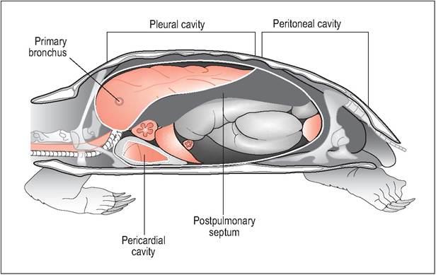
Figure 3.21 • Lateral view of chelonian (left lung, liver and stomach removed).
The heart occupies a cranial position because the lungs are restricted to the dorsal thorax.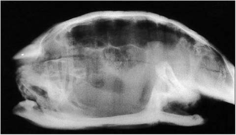
Figure 3.22 • Lateral radiograph (horizontal beam) showing lung fields.
Aquatic turtles
In aquatic species, respiration is aided by the hydrostatic pressure of water, which can draw air in and out of the lungs (Pough 2002). There are four groups of muscles involved in the respiratory cycle. Inspiration is created by the testo- coracoideus, which runs from the carapace to the medial scapula and dorsal coracoid, and the obliquus abdominis muscles, which help expand the cavity to create negative pressure. Expiration is via the diaphragmaticus and the transversus abdominis muscle, which compresses the celomic cavity (McCutcheon 1943; Wood & Lenfant 1976).
CLINICAL NOTE
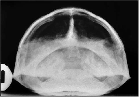
Figure 3.23 • Radiograph (horizontal beam) showing rostrocaudal view of lungs - the best view for assessing the lungs for pneumonia.
Chelonians cannot cough effectively as they lack a diaphragm. This factor combined with their huge lung volume and lack of a bronchi-ciliary transport system means they easily get pneumonia.
Accessory respiratory organs
Some semi-aquatic freshwater turtles possess the ability to absorb oxygen via well vascularized cloacal bursae, which they can use during periods of hibernation underwater. Others, like the softshelled turtles can remain submerged for hours in the mud, utilizing oxygen in the water by breathing through the skin and pharyngeal mucosa.