INTEGUMENT
Although it is commonly thought that reptiles have ‘slimy skin’ in fact, the converse is true as the skin is dry and has far fewer glands than either amphibians or mammals. It is also heavily keratinized with a lipid layer to prevent water loss.
The only glandular-type tissues are the femoral and precloacal pores seen in some lizards, which have a pheromonal function and are better developed in the male (Bellairs 1969d; Lillywhite & Maderson 1982).The epidermis is both thick and thin in order to form scales and it is these scales that make reptile skin a poor insulator of heat. Unlike fish scales, which can be scraped off, these scales are an integral part of the skin. The scales provide protection from abrasion, play a role in permeability, and tend to be thicker dorsally than ventrally. In some species they are developed into large plates and shields on the head. In snakes they are widened ventrally to form what are called gastropeges that are important for locomotion.
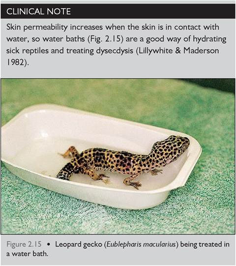
CLINICAL NOTE
Wound healing is slow in reptiles so stitches should be left in for at least 6 weeks (Bennett & Mader 1996; Rossi 1996). It is best to leave stitches in place until ecdysis occurs since the increased activity in the dermis and epidermis promotes better healing and strength.
Epidermis
The epidermis has three layers. The inner layer is called the stratum germinatum and consists of cuboidal cells that produce the protein keratin and the dividing cells of the intermediate layer. The intermediate layer has a lipid rich film that plays a major role in providing a water-permeable barrier in the skin. The outer stratum corneum is heavily keratinized into the scales. Two forms of keratin are produced in reptiles: alpha-keratin, which is flexible, and betakeratin, which provides strength and hardness and is unique to reptiles (Fig.
2.16). Beta-keratin is found in the scales of the chelonian shells whereas the alpha-keratin is found in the hinges or between the scutes (Bellairs 1969d; Harvey- Clark 1997; Lillywhite & Maderson 1982). It is at these weaker links that mites or infection like shell rot can be found (Harvey-Clark 1997).CLINICAL NOTE
The thick, keratinized skin of reptiles is at the expense of cutaneous sensation. Reptiles have far less sensory feeling in their skin than birds or mammals, which is why they are at more risk from thermal burns in captivity.
Dermis
The dermis consists of connective tissue, blood and lymphatic vessels, nerves, and pigment cells. In some species the dermis has bony plates called osteoderms. In chelonians this has fused with the vertebrae to form a shell.
Ecdysis
Ecdysis is the shedding of skin and is controlled by the thyroid gland. Changes in feeding behavior and activity
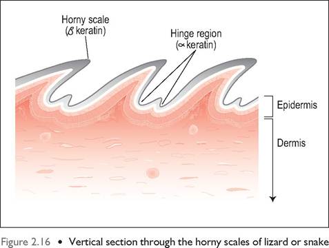
showing hinges between the scales. The hinges are made from the flexible alpha keratin while the beta keratin, which is unique to reptiles, gives strength and hardness to the scales.
occur prior to ecdysis and the reptiles become very susceptible to dehydration. Snakes tend to shed the whole skin, unlike lizards and chelonians which shed piecemeal, and this makes them even more vulnerable during ecdysis. In a healthy snake the whole process can take up to about 2 weeks.
During ecdysis the cells in the intermediate layer replicate to form a new three-layer epidermis. Once this process is complete, lymph diffuses into the area between the two layers and enzymes are released to form a cleavage zone. The old skin is shed and the new epithelium hardens, decreasing permeability to become the new skin (Harvey- Clark 1997; Lillywhite & Maderson 1982; Rossi 1996).
CLINICAL NOTE
During ecdysis the skin becomes more permeable and more vulnerable to parasites and infection.
Malnourished animals are hypoproteinemic and unable to produce enough enzymes to form a true cleavage zone, resulting in dysecdysis (failure to shed). Lack of moisture will also delay the process (Lillywhite & Maderson 1982).The production of color
Reptiles have pigment-containing cells called chromatophores that lie between the dermis and epidermis. These not only help in camouflage and sexual display but in thermoregulation. These pigment cells are not just confined to skin but can occur in the peritoneum in some species.
Melanophores produce the pigment melanin and lie deepest in the subepidermal layer. These melanin cells give rise to black, brown, yellow and gray coloration. Albinism in reptiles is caused by lack of melanin. The carotenoid cells are found beneath the epidermis above the melanophores and produce yellow, red and orange pigments (Bellairs 1969d).
Structural colors
The iridophores (guanophores) also lie in the dermis. These contain a semicrystalline product guanine (the breakdown product of uric acid) that reflects light. The blue wavelengths are reflected more to produce a blue color in an effect called Tyndall scattering. When combined with the yellow carotenoids this gives the color green, which is a common camouflage color in many reptiles (Bellairs 1969d).
Iridescence
Iridescence is caused by the physical properties of light on the thin and transparent outer layer of skin. When light strikes it from an angle the light spectrum is split into wavelengths of different colors. Depending on the color of the scales this will cause an iridescent effect when the snake moves. This feature is more obvious in black or dark snakes like the rainbow boa (Epicrates cenchria).
KEY POINTS
• The adrenal glands are intimately associated with the gonads in snakes and lizards.
• Reptiles have only one middle ear bone so their hearing is not acute like mammals.
• Jacobson’s organ is very well developed, especially in snakes, and plays a major role in olfaction.
• There is no consensual pupillary light reflex and the pupil is non responsive to atropine.
• Reptilian skin has few glands but has a lipid layer to prevent water loss.
• Sick animals suffer from dysecdysis due to lack of moisture and malnutrition.