Introduction
This book is written for Biology majors, nursing students, and pre-professional students whose curriculum requires a basic course in anatomy. An essential part of a course in mammalian anatomy should include a detailed dissection of a representative mammal.
Perhaps a human would be the most interesting for most students. However, since cadavers are not readily available and most institutions do not have the facilities to handle them properly, we feel that the domestic cat, Felis catus, is an extremely good substitute.Stephen O’Brien, a prominent scientist, involved in the HumanGenomeProject, recently pointed out that of the approximately 100 genes so far mapped in cats, each has its human counterpart. Furthermore, DNA base pair sequences are so similar that the genes and their arrangement can be determined when comparing the two. Even the gene position on the chromosomes is often very comparable (O'Brien, 1997). Perhaps, due to this genetic parallel, the anatomy of cats and humans is quite similar, with the exceptions largely related to differences in adaptations associated with food procurement and locomotion.
The differences between the cat and human provide interesting examples of adaptations among mammals. A number of systems in the cat exhibit adaptations for its predatory and carnivorous lifestyle—for example, the teeth are modified for feeding primarily on meat. The sharp canines pierce and hold struggling prey, the incisors nip small pieces from the meal, while a pair of lateral molariform teeth, called carnassial teeth, are designed to deal with tendons, ligaments, and shearing large chunks of meat. The cat’s predatory habits are further underscored by the general configuration of its head. The jaws are elongated to form a muzzle, which is an efficient shape for handling struggling prey. Another obvious suite of characteristics that exemplify both a predatory food habit and quadruped locomotion is illustrated in the structure of the fore- and hind-feet as well as the tail.
The cat walks on its tiptoes, thereby lengthening the leg and enabling it to run swiftly and function as an agile hunter. Additionally, the toes are equipped with laterally compressed, hooked, and retractile claws to climb and hold on to struggling meals. The long tail is used as a balance organ during its daily maneuvers and also as an organ of communication. If you have any doubts about the function of the tail, watch a cat as it leaps from a high perch or as it meets another cat or lies in wait for a mouse.Since humans are omnivorous and include a wide range of food in their diet, they exhibit less specialization of teeth. In the human, although canines, incisors, and molariform teeth are present, they are shaped differently and are adapted to handle a greater variety of foods, e.g., hamburgers, french fries, and salads at the salad bar. In contrast to cats, human molars are designed to grind, not to slice and cut, and since we are not predatory mammals, our jaw structure is shorter than the cat.
Human locomotion is bipedal which means that we walk upright on our hindlimbs and also on the entire foot from heel to toe. This permits the human foot to function as a stable gripping surface for locomotion. However, this stability does not allow us to develop into swift runners. Remember, most of our food doesn’t move too fast or require active capture! For this reason, in contrast to the cat, our finger and toe tips are protected by
dorso-ventrally flattened “claws ” called nails. Since human locomotion is normally bipedal and no longer requires the counterbalance of a rebounding rear end, the human tail has been greatly reduced in length and consists of a few vertebrae that are usually not externally obvious.
As the various systems are studied, major differences between the cat and the human will be noted.
★ ★ ★ ★ ★
To comprehend dissection directions and to become a literate anatomist, one needs to be able to understand and speak the language of anatomy. Students are expected to be able to communicate intelligently and precisely, both orally and in the written form.
In other words, you are expected to pronounce and spell anatomical and directional terms correctly. Directions for dissection in this book include the proper terminology. Therefore, it becomes imperative that the student recognizes these terms and becomes familiar with them. Our experience has indicated that students who read the text as well as consult the illustrations learn and understand mammalian anatomy far better than those who attempt to learn anatomy solely by “Picture book dissection!”The following discussion will provide you with necessary information to begin your study.
TERMS: ANATOMICAL AND DIRECTIONAL
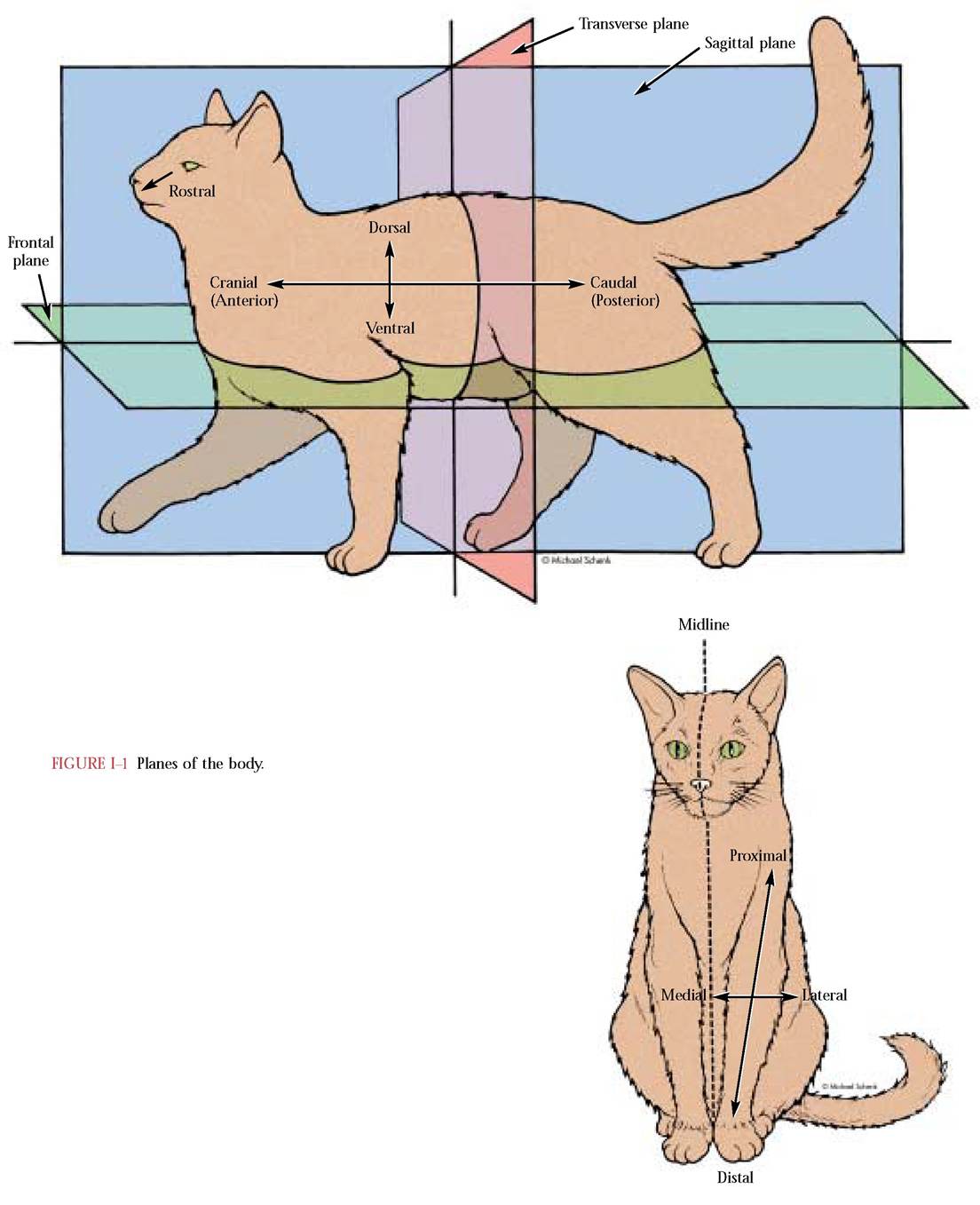
Some general directional terms include: Dorsal (the back or toward the back of the cat); Ventral (the belly or toward the belly of the cat); Cranial (toward the head); Caudal (toward the tail) [Figure I—1]. Just as often, in quadrupeds, Anterior (meaning ahead or before) and Posterior (meaning after or behind) will be encountered in descriptive anatomy. Very commonly, students will be directed toward the Medial (toward the midline) or Lateral (toward the side) aspect of the cat. The Midline is an imaginary line that extends directly down the middle of the dorsal and ventral surfaces. Frequently, you may encounter the directional terms, Proximal (next to or nearest to the point of origin or attachment) and Distal (some distance from the point of origin or attachment) [Figure I-1].
Often, planes of reference are important in understanding relationships of the morphology of organs, relationships among organs of a system within a body cavity, or relationships of organs and systems in a given view. In Figure I-1, a section parallel to the midline of the cat is referred to as
Sagittal. Therefore, there are an infinite number of sagittal sections, as long as you do not run out of cat. On the other hand, there is only one Mid-sagittal section which passes exactly down the midline of the body. A Transverse or cross section is illustrated in Figure I-1.
Just as there are an infinite number of sagittal sections, there are also an infinite number of transverse sections that may extend from the tip of the nose to the tip of the tail. Transverse sections are analogous to the slices of a loaf of bread, although usually much thinner. A Frontal section is made along the entire length of the cat parallel to the belly and back and illustrated in Figure I-1. Again, there can be numerous frontal sections.Suggested equipment check LIST
Dissection Tools
• 1 pair of fine point dissection scissors
• 1 scalpel handle, preferably No. 4, with replaceable blades
• Replaceable blades, preferably those designated as
21-25
• 1 steel probe, preferably a Huber-Mall
• 2 pair of straight forceps, one with medium points and the second with fine points
• Dissecting pins
Often, these tools are available in a kit, however, you will probably be advised by your instructor as to their purchase.
Other Equipment
• Safety goggles—these are strongly recommended to prevent eye injury from preservative fluids, bone chips and possible injury from dissection equipment
• Gloves—optional
• Lab jackets or coats—optional
• Sewing needles—these should be large needles
• Coat and button or carpet thread—this is heavy thread and should be white to prevent color from bleeding into the muscle
• Small spray bottle—any empty, clean spray bottle will work to hold preservative fluid to keep your specimen pliable and relatively fresh.