IRON METABOLISM
1. What is the oxidation state of the storage form of iron?
2. What is the oxidation state of iron for transfer across cell membranes?
3. What is the name of the transport form of iron?
4.
Would iron in its transport form be toxic? If not, why not?5. What are the normal limitations to iron absorption? Can iron toxicity occur as a result of excess ingestion and subsequent absorption?
Free iron (Fe3+) catalyzes the separation of free radicals from molecular oxygen, and oxygen free radicals are toxic. To avoid toxicity, intracellular iron is either bound to or incorporated into various proteins. It is transported and stored in the protein-bound form in its ferric (Fe3+) oxidation state. To be transported across membranes, iron must be in its ferrous (Fe2+) oxidation state.
A large proportion of ingested iron is reduced to ferrous iron (Fe2+) in the stomach. Within the duodenum and jejunum, most of the ferrous iron is absorbed into the intestinal epithelial cells. Iron absorption, transport, storage, and usage are summarized in Figure 3-10. From the intestinal cell, it enters the blood or can combine with a cellular protein (apoferritin) to become ferritin, a storage form of iron. Within 2 or 3 days, the ferritin is either converted back to its free form (Fe2+) and absorbed into the blood or is cast into the intestinal lumen. The latter situation would be a result of the normal turnover of intestinal epithelial cells as they migrate from the crypts to the tips of the villi, from which they are exfoliated (shed into the lumen). The iron that enters the blood combines with apotransferrin (a plasma protein) to form transferrin. Combining with a protein prevents it from being excreted by the kidneys (the combination is poorly filtered at the glomerulus).
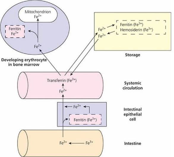
■ FIGURE 3-10 Summary of iron absorption, storage, and use.
Iron must be in the ferrous (Fe2+) oxidation state to be transported across membranes. Intracellular iron is bound to or incorporated into various proteins or other chelates in its ferric (Fe3+) oxidation state to reduce its toxicity because free iron can catalyze free radicals from molecular oxygen and hydrogen ions and can have disastrous consequences for biological materials. Transported iron is bound to the protein apotransferrin and is known as transferrin. Iron is stored in tissues as either a diffuse, soluble, mobile fraction (ferritin) or as insoluble, aggregated deposits (hemosiderin). Principal locations of iron storage are the liver and spleen, followed by the kidney, heart, skeletal muscle, and brain. In the bone marrow, all erythroid forms have surface membrane receptors for transferrin. When internalized, released iron is transported into the mitochondria of developing erythrocytes, where it is incorporated into the heme molecule or it combines with the protein apoferritin to be stored as ferritin. (From Reece WO, Swenson MJ. The composition and functions of blood. In: Reece WO, ed. Dukes’ Physiology of Domestic Animals. 13th edn. Ames, IA: Wiley-Blackwell, 2015.)Within the bone marrow, all the erythroid forms, including reticulocytes, have surface membrane receptors for transferrin. Plasma transferrin binds to these receptors, becomes internalized by endocytosis, and releases its iron, and the apotransferrin is returned to the plasma.The internalized iron is either transported into the mitochondria of the developing erythrocyte, where it is incorporated into the heme molecule, or it combines with apoferritin to be stored as ferritin in its ferric (Fe2+) oxidation state.
Two factors generally affect the absorption of iron from the intestinal epithelium into the blood: (1) the extent of the iron stores in the body and (2) the rate of erythropoiesis. If the requirement for iron increases and the iron stores are empty, absorption increases. If the requirement for iron decreases and the iron stores are adequate, absorption of iron from the intestine decreases. It seems that there is a self-limiting mechanism for iron absorption based on need. However, excess iron can be ingested and subsequently absorbed, inducing iron toxicity. The excretion of iron is minimal, so that regulation is unidirectional, i.e., controlled absorption. Iron with transferrin can be released to tissue cells anywhere so that excess iron can be deposited in all cells, especially those of the liver. Ferritin is a storage form of iron (see previous text). In addition, a more insoluble form, hemosiderin, accumulates in times of excess. The liver is the principal organ for iron storage. When liver stores are adequate the production of apotransferrin decreases and when they are depleted the production of apotransferrin increases. Animals with iron-deficiency anemia have high concentrations of apotransferrin.