JOINTS AND SYNOVIAL FLUID
1. What is another name for the connection between component parts of the skeleton that is otherwise known as a joint?
2. What is the term used to describe inflammation of a joint?
3.
What facilitates the gliding of two surfaces of a synovial joint over each other?4. What is a joint capsule?
5. What part of the joint capsule secretes synovial fluid?
6. Do joints have a lymph drainage?
7. What functions are served by nerves that supply joints?
8. Are there pain nerve fibers.in articular cartilage? What.is the distribution of pain. nerve fibers that are associated with a joint?
9. Does the synovial membrane cover the articular cartilage?
0. What are synoviocytes?
11. What are the chief functions of synovial fluid?
2. What component of synovial fluid provides for its viscosity?
3. What is the difference in viscosity of synovial fluid among joints of different sizes?
4. Are normal plasma constituents common to synovial fluid?
L5. Describe adult articular cartilage. Does it have cells, blood vessels, and a nerve supply?
6. What provides the growth zone for endochondral ossification of the epiphysis?
L7. How does intermittent pressure on articular cartilage relate to its nutrition?
8. What substances in synovial fluid contribute to its lubricating properties?
9. How does compression on articular cartilages contribute to lubrication?
0. What is weeping lubrication?
The connection between any of the skeleton’s rigid component parts is known as a joint. These connections are also described as articulations. The study of joints is termed arthrology and inflammation of joints is referred to as arthritis. Arthritis is a common malady among domestic animals; therefore, this brief study of the anatomy and physiology of joints is intended to assist students’ understanding of joint diseases. A slightly movable joint was described for the connection of contiguous vertebrae (see Figure 7-5).
Synovial joints are those that allow one surface to glide over another (Figure 7-21). This motion is facilitated by the presence.of articular cartilage on each bone surface of the articulation and also by the presence,of synovial fluid. The synovial joint is enclosed by a joint capsule. Synovial fluid is contained within the cavity of the joint capsule and is secreted by its inner membrane, the synovial membrane. The outer layer of the joint capsule is a fibrous layer that extends from the periosteum of each bone and contributes to the stability of the joint. A few joints contain a meniscus (fibrocartilaginous plate) that serves a cushioning function.
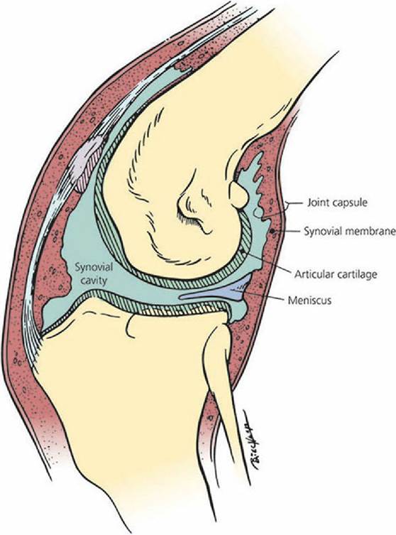
■ FIGURE 7-21 Articular cartilage covers the opposing bony surfaces of a synovial joint as shown in this diagram of a stifle. The joint space is filled with synovial fluid from the synovial membrane of the surrounding joint capsule. A meniscus composed of Iibrocartilage extends into the joint cavity. (From Dellmann HD and Eurell JA, eds. Textbook of Veterinary Histology. 5th edn. Baltimore, MD: Williams & Wilkins, 1998.)
Blood, Lymph, and Nerve Supply of Joints
The blood and nerve supply of a synovial joint is shown in Figure 7-22. The arteries that supply a joint and adjacent bone generally have a common origin. These arteries usually enter the bone near the line of capsule attachment and form a network around the joint. Capillaries from this network are one of the sources of nutrition to articular cartilage that was noted in the previous section. Lymph vessels are present with blood vessels and the lymph vessels that leave a joint drain into regional lymph nodes. Diffusion between the joint cavity and the blood and lymph capillaries takes place readily.
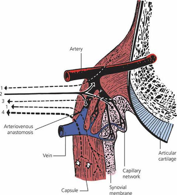
■ FIGURE 7-22 Blood and nerve supply of a synovial joint.
An artery is shown supplying the epiphysis, joint capsule, and synovial membrane. Note the arteriovenous anastomosis. The articular nerve contains the following: (1) sensory fibers (mostly pain) from the capsule and synovial membrane, (2) autonomic fibers (postganglionic, sympathetic to blood vessels), (3) sensory fibers (pain, and others with unknown functions) from the adventitia of blood vessels, and (4) proprioceptive fibers from Ruffini endings and from small lamellated corpuscles (not shown). Arrows indicate the direction of conduction. (From Gardner E, Gray DJ, O’Rahilley R. Anatomy. 4th edn. Philadelphia, PA: WB Saunders, 1975.)Nerve supply to a joint has two principal functions. The first function has to do with pain and reflex responses that may accompany joint disease. The second function is associated with their role in posture, locomotion, and kinesthesia, which is a sense mediated by stimulation of end organs in muscles, tendons, and joints in response to body movements and tension (see Chapters 4 and 5). The pain fibers are distributed within the fibrous layer and synovial membrane of the joint capsule.
Synovial Membrane
The synovial membrane is a vascular connective tissue that lines the inner surface of the joint capsule but does not cover the bearing surfaces (the articular cartilage). Synoviocytes within the synovial membrane synthesize synovial fluid by an active, energy-requiring process.
The chief functions of synovial fluid are joint lubrication and nourishment of the articular cartilage. Synovial fluid is a sticky, viscous fluid, often like egg white in consistency. It is usually slightly alkaline and ranges from colorless to deep yellow. The color and viscosity vary with species and type of joint. Fluid from large joints is usually less viscous than that from small joints. The viscosity of synovial fluid is attributable almost entirely to hyaluronic acid. Other chemical constituents of synovial fluid are those that are normally present in blood plasma.
Synovial fluid normally contains a few cells that are mostly mononuclear. Examination of the cellular and chemical content and physical characteristics can be a valuable diagnostic aid when evaluating joint disease.Articular Cartilage
Adult articular cartilage is usually hyaline in nature, avascular, and aneural, and has an acellular matrix that surrounds a relatively small number of cells called chondrocytes. It is a highly specialized connective tissue with biochemical and biophysical characteristics that enable it to play a dual role as a shock absorber and as a bearing surface. During the growth period, articular cartilage provides the growth zone for endochondral ossification in the epiphysis. During growth, articular cartilage is capable of regeneration and thus repairs defects that may arise. However, when growth ceases, it loses much of its power of repair. Cartilage is a resilient and elastic tissue that becomes thinner when compressed and slowly regains its original thickness when the pressure is released. Intermittent pressure associated with compression and release of compression causes cartilage to thicken by taking up fluid. Synovial fluid is absorbed by this sponge-like property and diffuses through the cartilage matrix to provide for its nutrition. Other possible sources of nourishment for articular cartilage include diffusion from epiphyseal vessels that loop through subchondral bone and diffusion of fluid from capillaries associated with the arterial circle around the joint at the line of capsular attachment.
Lubrication of Synovial Joints
The fluids that lubricate a synovial joint are the synovial fluid and fluid pressed from articular cartilage during compression.,Substances within synovial fluid that contribute to its lubricating properties are hyaluronic acid and a glycoprotein known as lubricin. Both of these substances are secreted by the synovial membrane and lubricate the articular surface during light loads associated with minimal articular cartilage compression. During heavy loads, the synovial membrane fluids are displaced from the articular cartilages and the compression causes fluid from the cartilage to be expressed and form a layer between the opposing surfaces. The lubrication provided by the cartilage fluid is known as weeping lubrication. Articular cartilage has been compared with a stiff sponge; it resists tensile stresses, shows elastic deformation under load, contains a high proportion of extracellular fluid (hyperhydrated), and exudes fluid under pressure (which is of major importance in lubrication).
■