BONE REPAIR
1. What happens to osteocytes, the periosteum, and bone marrow when the blood supply is disrupted after bone fracture?
2. Will bone repair occur if a blood supply is not restored to a fracture site?
3.
As related to bone repair, what.is a callus?4. What is the source of the osteogenic cells for the external and internal callus?
5. What determines whether a callus will be composed of spongy bone or cartilage?
6. What eventually happens to a cartilage callus?
7. Will spongy bone be replaced.by compact bone at the fracture site?
8. What determines when.remodeling of initial bone repair will occur?
Bone fractures are the most common consequences of bone injury. Fractures can result in separation of bone parts with loss of alignment, separation of periosteum and endosteum, and severe bleeding followed by clot formation. The torn blood vessels can be those that supply Volkmann canals, Haversian systems, and the periosteum and endosteum at the fracture site. In the vicinity of the disrupted blood supply, the osteocytes begin to die and the periosteum and bone marrow become necrotic. The acute inflammatory condition that follows brings phagocytic cells into the area for clearance of blood clot components and necrotic tissue. New blood vessels enter the damaged area, and new bone formation begins. Bone formation does not occur until a blood supply has been established.
The most common type of bone repair involves the formation of a callus. This type takes place when the broken ends are not perfectly realigned and stabilized. A collar of repair tissue forms around the external surface of each broken end, and when a bridge is formed across the break, it is known as the external callus. The healthy intact periosteum is the source of the osteogenic cells for the external callus, whereas endosteum is the source for the internal callus.
Depending on the richness of the periosteal capillaries, the callus will be composed of either spongy bone or cartilage. Inadequate blood supply predisposes to cartilage formation. When cartilage is formed, it is subsequently replaced by bone. The transformation from cartilage to bone is similar to that previously described for growth of long bones from the epiphyseal plate. The chondrocytes hypertrophy and the cartilage matrix becomes calcified. The calcified cartilage is removed and replaced with spongy bone after the entrance of blood vessels. Any dead bone that was incorporated into the callus is removed by the action of osteoclasts and is replaced by bone formed by osteoblasts that move into the spaces created by osteoclastic activity. As compact bone is formed at the fracture site, the spongy bone in the periphery of the callus is no longer needed to provide strength, and therefore it is resorbed. Final remodeling occurs when stresses associated with normal use return. A summary of fracture healing is shown in Figure 7-20.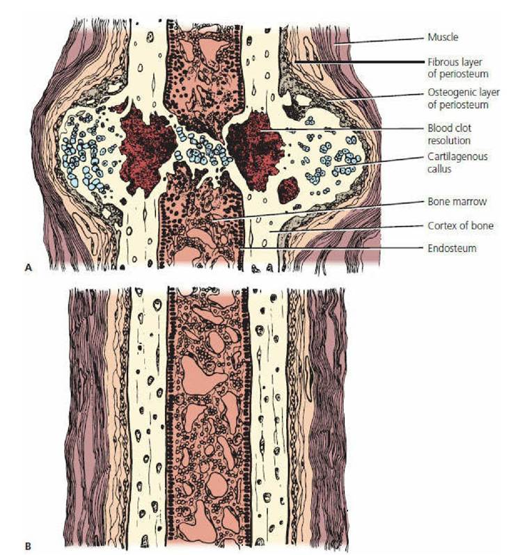
■ FIGURE 7-20 Bone fracture repair. A. Fracture has been reduced and immobilized. Repair involves the appearance of a palpable callus. A cartilaginous callus precedes the mineralized callus. B. Fracture completely healed. The bone has been remodeled to conform to lines of stress. The eriginal fracture site is obliterated. (From Whittick WG. Canine orthopedics. 2nd Edn. Philadelphia, PA: Lea & Febiger, 1990.)
■