» Lymphatic Structures of the Thorax (see also pp. 245247)
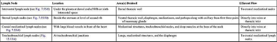
The lymph nodes of the thorax are summarized in Table 13.2. The thin-walled thoracic duct begins between the crura of the diaphragm as the continuation of the cisterna chyli. It accompanies the aorta and azygos vein forward and, level with the heart, passes obliquely to the left, crossing the esophagus, to gain a position within the left side of the cranial mediastinum.
It follows the esophagus to the thoracic inlet, where it opens into one or other of the larger veins. However, occasionally it ends more caudally by joining the azygos vein or even opening into one of the mediastinal lymph nodes. The duct, which has a diameter of 2 to 3 mm in a medium-sized dog, may be plexiform (see Fig. 7.57). Within the chest it receives additional lymph from various thoracic structures and nodes of the left side; a separate right lymphatic duct provides similar drainage for structures of the right side. One or both commonly receive the corresponding tracheal duct(s). In cats the thoracic duct courses from the left dorsal aspect of the aorta to terminate in the left jugular vein. In both species the thoracic duct may have multiple collaterals.» TABLE 13.2
Thoracic lymph nodes
Comprehension Check
Develop an understanding of projections of the lungs and the heart on the chest wall, and use that information for cardiac and pulmonary auscultation.
More on the topic » Lymphatic Structures of the Thorax (see also pp. 245247):
-
Veterinarian -