LYMPHATIC STRUCTURES OF THE THORAX (See also pp. 259-260.)
A single intercostal lymph node may be present under the pleura at the dorsal end of the fifth or sixth intercostal space. It drains the structures of the dorsal thoracic wall and sends its efferent vessels to the cranial mediastinal nodes (Figure 7-55/6).
The sternal lymph nodes are large—up to 2 cm in length—and lie embedded in fat beside the sternum at
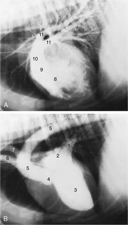
Figure 13-21 Contrast medium in the canine right (A) and left (B) ventricles marking the great vessels. The catheter is in the cranial vena cava. 1, Pulmonary veins; 2, left atrium; 3, left ventricle; 4, position of aortic valve; 5, aorta; 6, brachiocephalic trunk; 7, left subclavian artery; 8, right ventricle; 9, position of pulmonary valve; 10, pulmonary trunk; 11, pulmonary arteries.
the level of the second rib. They receive lymph from the muscles of the ventral thoracic wall, the diaphragm, and the mediastinum and may collaborate with the axillary lymph nodes in draining the first three pairs of mammary glands. Their efferent vessels go to veins at the thoracic inlet (Figure 7-55/10).
The cranial mediastinal lymph nodes are variously related to the large blood vessels in front of the heart. They drain structures in the mediastinum (including the tracheobronchial nodes) and the deep muscles at the base of the neck. Their outflow also enters the veins at the thoracic inlet (Figure 7-55/8).
The tracheobronchial lymph nodes (Figure 13-13, A) are scattered about the termination of the trachea and the principal bronchi. They drain the lungs as well as mediastinal structures and part of the diaphragm. Their efferent vessels pass to the cranial mediastinal nodes.
The thin-walled thoracic duct begins between the crura of the diaphragm as the continuation of the cisterna chyli. It accompanies the aorta and azygous vein forward and, level with the heart, passes obliquely to the left, crossing the esophagus, to gain a position within the left side of the cranial mediastinum. It follows the esophagus to the thoracic inlet, where it opens into one or other of the larger veins; occasionally it ends more caudally, joining the azygous vein or even opening into one of the mediastinal lymph nodes. The duct, which has a diameter of 2 to 3 mm in a medium-sized dog, may be plexiform (Figure 7-57). Within the chest it receives additional lymph from various thoracic structures and nodes of the left side; a separate right lymphatic duct provides similar drainage for structures of the right side. One or both commonly receive the corresponding tracheal duct(s). In cats the thoracic duct courses from the left dorsal aspect of the aorta to terminate in the left jugular vein. In both species the thoracic duct may have multiple collaterals.