MALE
Testes
The ferret scrotum has two cavities, each of which houses a testis and epididymis (Figs. 12.18 ad 12.19a, b). The epididymis is composed of a mass of convoluted spermatic duct, which is divided into head, body, and tail on the dorsal aspect of the testis as it lies in the scrotal sac.
The head of the epididymis receives the efferent ductules as they leave the testis at the cranial pole. The body of the epididymis has the main mass of the convoluting spermatic duct, which passes to the tail of the epididymis at the caudal pole of the testis. The spermatic duct becomes apparent as a simple duct to the tail of the epididymis and passes cranially on the dorsal aspect of the testes as the ductus deferens (vas deferens). It is medial to the body of the epididymis.The ductus deferens is accompanied by the deferent artery and vein and the main testicular artery and vein, which, with the nerve and lymphatic system vessels, form the spermatic cord. It passes cranially to the inguinal passage into the abdomen where it then parts company with the testicular artery, looping over the urethra through the prostate gland.
Histologically, the testis, blood vessels, nerves, lymphatics, and ductus deferens are wrapped in a pouch of peritoneum called the vaginal sac. This sac extends through the inguinal canal in development prior to the descent of the testis.
Castration of hob ferrets is a common operation (Bennett & Pye 2000) but a more delicate and equally important operation is that of vasectomy (Lewington 2003c). The treated hob can then be used to take jills off heat, to rest them from breeding without recourse to hormonal injection.
During vasectomy surgery the common vaginal tunic of the spermatic cord must be delicately dissected open to expose the ductus deferens and avoid injury to the main testicular artery. The ductus deferens lies medial to the testicular artery and vein inside the spermatic cord (Lewington 2003g).
The hob genitalia resemble the dog in having an os penis or baculum. The os penis of the ferret, and indeed other mustelids, is a bony strengthening rod, as in the dog, but has an exterior curled point that makes urethral catheterization difficult. Despite the presence of the curled end, catheterization can be achieved using a 3.5 French catheter (Lucas 2000) (Fig. 12.20).
Prostate gland
The prostate is a glandular structure, which adds elements to the seminal fluid. It lies at the base of the bladder surrounding the urethra (Fig. 12.18). It is not very distinct in the young ferret. As it completely surrounds the urethra, inflammation of the gland will affect the urinary outflow. At the level of the prostate the ductus deferens from each side opens into the urethra.
Small mammals
249
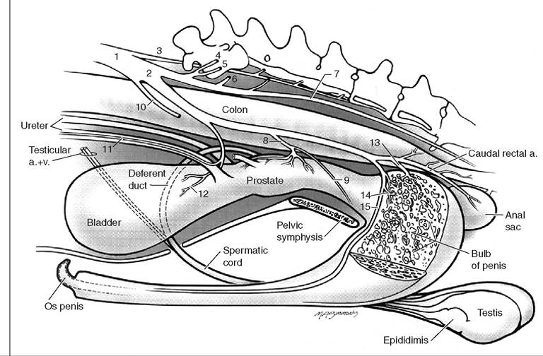
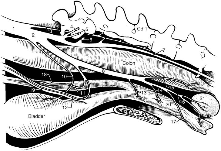
1. Internal iliac artery
2. Internal pudendal artery
3. Medial sacral artery
4. Iliolumbar artery
5. Cranial gluteal artery
6. Caudal gluteal artery
7. Lateral caudal artery
8. Prostatic artery
9. Urethral artery
10. Umbilical artery
11. Ureteral artery branch
12. Caudal vesicle artery
13. Artery of the bulb
14. Deep artery of penis
15. Dorsal artery of penis
Figure 12.18 • The bladder in relation to the hob reproductive system. (Courtesy of Lippincott Williams & Wilkins and Howard Evans.)
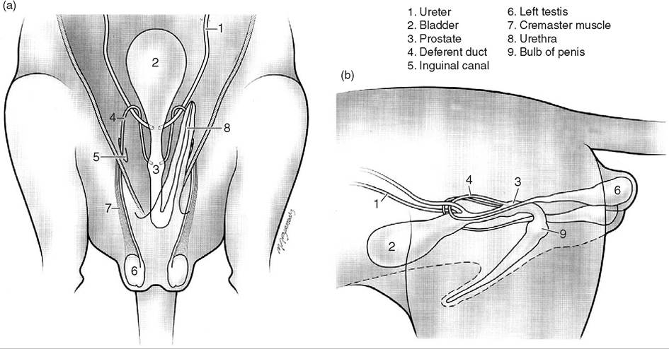
Figure 12.19 • (a) Hob genitalia ventral view; (b) Hob genitalia right lateral view. (Courtesy of Lippincott Williams & Wilkins and Howard Evans.)
Prostatic cysts can develop in association with adrenal gland neoplasia (AGN), which can cause urethral obstruction (Fig. 12.21). The gland becomes enlarged in mostly American sterilized hobs as a frequent complication of
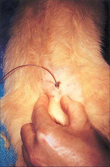
Figure 12.20 • Urethral catheterization of the hob ferret.
Note the J-shaped os penis to the top right of the catheter. The urethra does not bend around inside the os penis. Slide courtesy of Dr. Anthony Lucas.AGN. This is not a problem of unsterilized hobs or animals sterilized at a mature age but is common in American male ferrets, which are sterilized at 6 weeks (Jenkins & Brown 1993). Once adrenalectomy is carried out the prostate rapidly reduces in one to two days. Surgery can be performed on this condition (Bennett & Pye 2000).
Female
Ovaries
The weight of an ovary is 94-183 mg in a 600-800 g ferret, with the average size being 0.45 cm long by 0.55 cm wide
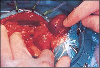
Figure 12.21 • Prostatic cyst in a ferret. The large prostatic cyst can be seen dorsal to the reflected and elevated bladder. Note the use of a stay retractor to facilitate exposure of the abdomen. The hooks of the stays have been placed in the muscle of the body wall. Slide courtesy of Prof. Avery Bennett.
1. Internal iliac artery
2. Internal pudendal artery
3. Medial sacral artery
4. Iliolumbar artery
5. Cranial gluteal artery
6. Caudal gluteal artery
7. Lateral caudal artery
8. Vaginal artery
9. Uterine horn
10. Umbilical artery
11. Uterine artery
12. Ureteral branch
13. Urethral artery
14. Artery to vestibular bulb
15. Caudal rectal artery
16. Perineal artery
17. Artery of the clitoris
18. Uterine horn
19. Ureter
20. Vagina
21. Anal sac
Figure 12.22 • The bladder in relation to the jill reproductive system. Courtesy of Howard Evans.
and 0.21 cm thick (Evans & An 1998). When mature, the left ovary lies caudal to the middle of the 14 th rib and caudal to the left kidney. The right ovary is caudal to the middle of the last rib (15th) and also caudal to the right kidney.
The ovaries are suspended by ligaments from the abdominal wall.Uterus
The ferret uterus is bicornuate, comprising two long, tapering uterine horns that combine immediately in front of the cervix to form a short uterine body (Fig. 12.22). The mature uterine horns are about 4.3 cm long and 0.22 cm wide, while the body is 1.7 cm long and 0.25 cm wide (Evans & An 1998). Blood supply is by the companion ovarian and uterine arteries and veins (Fig. 12.25); sympathetic innervation comes from the aortic and renal plexuses.
Ovariohysterectomy is done as per the cat. The ovarian ligaments are loose and easily torn to exteriorize the ovaries.
CLINICAL NOTE
During spaying, the ovaries might well be obscured by the presence of fat and thus care must be taken in making sure the full ovary is removed, and no remnants are left, as these can produce hormonal problems.
Estrous cycle
The sexual cycle of the ferret comes with maturity at 6-9 months of age. Breeding is tuned to the increasing daylight periods in spring. Normally the vulva is hardly visible in the live animal, becoming red and swollen when the jill is in heat.
Ovulation is stimulated by coitus, with the jill becoming anestrus 1 week after fertilization. Failure of the jill to mate results in continuous hormonal stimulation of estrogen from the ovaries, leading to the dangerous condition of hyper- estrogenism. The condition arising is postestrus anemia. The jill's vulva continues to be swollen, she becomes anemic, shows alopecia and is very sick (Fig. 12.23). The high blood estrogen from the active ovaries causes suppression of the bone marrow and a non-regenerative anemia with depression of PCV below 25%. If the PCV falls below 15% it is considered irreversible. Treatment is by immediate ovariohysterectomy if the PCV is still high or else blood transfusion and intensive care is required. In addition the uterus can develop pyometra as a result of ascending vaginal infections or as a sequel to postestrus anemia.
Thus the female ferret, unlike other mammals, must be mated, taken out of estrus by a vasectomized hob, or given chemical estrus suppression drugs (Lewington 2003c). Feral ferrets and other Mustelidae are usually kept mated in the season or die quickly. American pet ferrets do not get postestrus anemia as non-breeding jills are sterilized at 6 weeks of age; however they are plagued with adrenal gland neoplasia (AGN) and insulinoma, as shown later.
KEY NOTES
• Prostatitis occurs in the sterilized male, associated with adrenal gland neoplasia (AGN).
• The os penis has a curled end making catheterization difficult but not impossible.
• Postestrus anemia occurs in the unmated female during estrus.
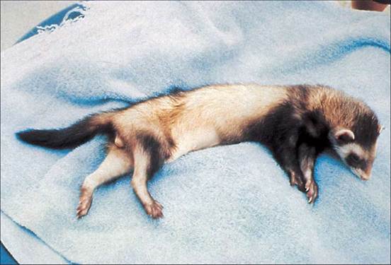
Figure 12.23 • Jill with postestrus anemia showing alopecia and swollen vulva. Slide courtesy of D. Manning, University of Wisconsin.