MAMMARY GLANDS
The mammary glands contribute to the contours during pregnancy and lactation. Dogs generally have five pairs of mammary glands, spread along the ventral aspect of the trunk (Figure 10-31, C, and Figure 10-32, B-C).
The two cranial pairs are thoracic, the next two abdominal, and the caudal-most pair inguinal in position. A distinct midline separation is noted between the left and right mammary chains. Their pattern is often staggered, which is a favorable arrangement because it makes all teats equally accessible to the pups when the bitch suckles lying on her side. The glands are very small in the virgin (with the teats hidden by hair) but become very swollen, pendulous, and confluent with their ipsilateral neighbors toward parturition and during lactation. They regress greatly in the parous but nonpregnant and nonlactating bitch, although the teats remain enlarged in parous bitches, in which they are superimposed on the abdominal organs in ventrodorsal radiographs. The teats, which occur in rudimentary form in males, are bare and perforated at their tips by 10 or 12 fine openings through which milk is drawn.The cat has four pairs of mammary glands of which the teat is situated approximately 3 cm from the midline. The nonlactating teat of the cat is about 1 mm high, perforated by four to eight openings, and enlarges almost tenfold in lactation.
In dogs and cats, the blood supply to the mammary glands varies in detail but mainly originates from the lateral and internal thoracic and the external pudendal arteries; some assistance may be provided by lesser vessels from other sources. In most cases, the cranial three (in cats two) glands are supplied craniolaterally by the lateral thoracic artery (from the axillary) and deeply by the cranial superficial epigastric artery and perforating branches of the intercostal arteries (both from the internal thoracic).
The two caudal pairs receive blood from the caudal superficial epigastric artery (from the external pudendal) and deeply from branches of the cranial abdominal and deep circumflex iliac arteries. The veins are satellite. Both arteries and veins anastomose freely, forming arterial and venous plexuses (Figure 14-2, A), which may cross the midline.Lymph from the cranial three (in cats two) glands goes to the axillary, accessory axillary, and sternal nodes, and that from the two caudal glands goes to the superficial inguinal (mammary) node, which is located dorsal to the caudal border of the inguinal mammary gland (or in the cat, also to the caudal epigastric nodes, which are small and located along the course of the caudal epigastric vessels). In the dog, the third pair usually drains toward the axillary lymph node but may also drain caudally. The pathways are erratic, and some
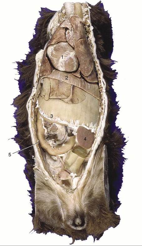
Figure 14-1 Ventral view of a canine trunk, demonstrating the so-called intrathoracic part of the abdomen. 1, Heart; 2, diaphragm; 3, distended stomach (with attachment of greater omentum); 4, spleen; 5, duodenum.
lymph may cross the midline. It is said that in the cat the lymph vessels do not cross the midline nor penetrate the thoracic wall. The superficial inguinal nodes and caudal glands are related to the vaginal process, which is vulnerable during surgical removal of a diseased gland; injury to the process may cause inadvertent opening of the peritoneal cavity. In both species the superficial inguinal nodes drain the adjacent part of the abdominal wall in addition to the caudal mammary glands.
These details obtain importance from the prevalence of mammary tumors in both dogs and cats. In bitches they are the commonest of all tumors and show a disturbingly high (ca. 50%) incidence of malignancy. Although somewhat less common in cats, mammary
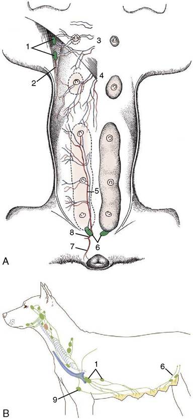
Figure 14-2 Blood vessels and lymphatics of the canine mammary glands. A, Ventral view of the mammary glands, blood vessels, and certain lymph nodes. B, Lateral view of regional lymph nodes. 1, Axillary and accessory axillary lymph nodes; 2, branch of lateral thoracic artery; 3, perforating branches of internal thoracic vessels; 4, branches of the cranial superficial epigastric vessels; 5, caudal superficial epigastric artery; 6, superficial inguinal lymph nodes; 7, ventral labial branch to vulva; 8, external pudendal artery; 9, sternal lymph node.
tumors are even more likely to be malignant in this species.