ŧ Mammary Glands
The mammary glands contribute to the contours during pregnancy and lactation. Dogs generally have five pairs of mammary glands and the cat has four pairs, spread along the ventral aspect of the trunk (see Figs.
10.31C and 10.32B and C). The two cranial pairs are thoracic, the next two abdominal, and the caudalmost pair inguinal in position. A distinct midline separation is noted between the left and right mammary chains. The often staggered pattern makes all teats equally accessible to the pups when the bitch suckles lying on her side. The glands are very small in the virgin (with the teats hidden by hair) but become very swollen, pendulous, and confluent with their ipsilateral neighbors toward parturition and during lactation. The size increase is nearly 10-fold in the cat. They regress greatly in the parous but nonpregnant and nonlactating bitch. The teats, which occur in rudimentary form in males, are bare and perforated at their tips by 10 or 12 fine openings in the dog and 4 to 8 openings in the cat.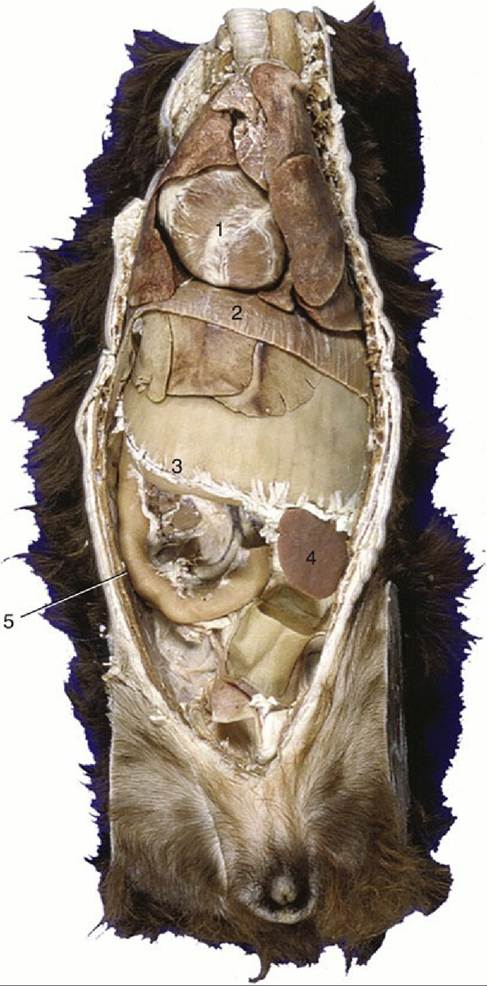
FIG. 14.1 Ventral view of a canine trunk, demonstrating the so-called intrathoracic part of the abdomen. 1, Heart; 2, diaphragm; 3, distended stomach (with attachment of greater omentum); 4, spleen; 5, duodenum.
In dogs and cats, the lateral and internal thoracic and external pudendal arteries are the major sources of blood supply to the mammary glands. The veins are satellite. Both arteries and veins anastomose freely, forming arterial and venous plexuses (Fig. 14.2A), which may cross the midline.
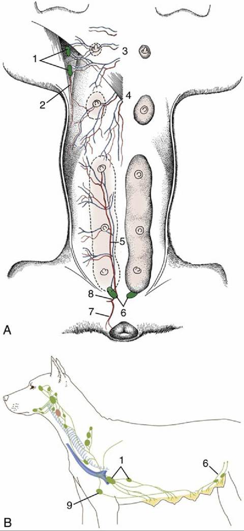
FIG. 14.2 Blood vessels (red) and lymphatics (green) of the canine mammary glands. (A) Ventral view of the mammary glands, blood vessels, and certain lymph nodes.
(B) Lateral view of regional lymph nodes. 1, Axillary and accessory axillary lymph nodes; 2, branch of lateral thoracic artery; 3, perforating branches of internal thoracic vessels; 4, branches of the cranial superficial epigastric vessels; 5, caudal superficial epigastric artery; 6, superficial inguinal lymph nodes; 7, ventral labial branch to vulva; 8, external pudendal artery; 9, sternal lymph node.The lymph drainage pathways are erratic, and some lymph may cross the midline. It is said that in the cat the lymph vessels do not cross the midline nor penetrate the thoracic wall (Table 14.1). The superficial inguinal nodes and caudal glands are related to the vaginal process, which is vulnerable during surgical removal of a diseased gland; injury to the process may cause inadvertent opening of the peritoneal cavity. In both species the superficial inguinal nodes drain the adjacent part of the abdominal wall in addition to the caudal mammary glands. These details of lymph drainage are important because of the high prevalence of mammary tumors in both dogs and cats. In bitches they are the commonest of all tumors and show a disturbingly high (ca. 50%) incidence of malignancy. Although somewhat less common in cats, mammary tumors are even more likely to be malignant in this species.