Mammary Glands of the Cow
The udder of the cow comprises four individual glands, referred to as quarters. The skin of the udder is covered with fine hair; however, the teat is completely hairless. The right and left halves of the udder each consist of a cranial (front) quarter and a caudal (hind) quarter.
Each side of the udder is almost completely independent of the other insofar as blood supply, nerve supply, and suspensory apparatus are concerned (discussed later).Ventrally, the two halves of the udder are demarcated by a longitudinal furrow, the intermammary groove, which corresponds to a median septum of connective tissue dividing left and right halves. Because of the relative isolation of each side, half of the udder can be removed surgically without damaging the other half, as might be done to treat an aggressive tumor. The two quarters in each half are separate from one another as far as the gland tissue and duct system are concerned. Thus, all the milk from one teat is produced by the glandular tissue of that quarter. The vasculature, nerve supply, and lymphatic drainage, however, are common to both quarters of a given half.
The parenchyma of the lactating mammary gland consists of secretory tissue and the ducts of the gland (Fig. 29-1). The secretory units, the alveoli, are lined by a simple epithelium that varies from columnar to cuboidal in height. The alveoli are the chief structures for actual milk production, although the initial portion of the associated duct is also lined with secretory epithelium.
The various small initial ducts converge to form larger ducts, and these converge to form yet larger ones, all of which eventually terminate in a large single basin, the lactiferous sinus. The lactiferous sinus is sometimes described as being divided into a large cavity within the quarter itself, the pars glandularis (gland cistern), and a smaller cavity within the associated teat called the pars papillaris (teat cistern) (Fig.
29-1). The demarcation between gland cistern and teat cistern frequently is marked by a circular ridge (annulus) that contains a vein and some smooth muscle fibers.The wall of the empty cistern contains numerous overlapping longitudinal and circular folds that are obliterated through expansion of the wall when it is full of milk. There may also be diverticula (pockets) in the wall of the gland cistern.
The teat cistern is continuous with the exterior of the teat through a narrow opening in the end of the teat, the papillary duct (commonly called streak canal or teat canal), which opens at the ostium papillae. The bovine streak canal is about 8.5 mm long, and its lumen is normally closed by epithelial folds that project inward from the wall of the streak canal, leaving only a star-shaped potential opening. A sphincter of smooth muscle fibers surrounds the streak canal at the distal end of the teat.
Suspensory Apparatus
The udder of a lactating dairy cow can weigh as much as 60 kg, so the organ is supported by
BOVINE UDDER
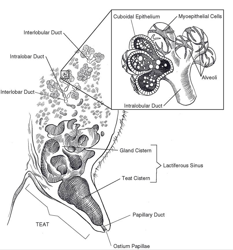
Figure 29-1. Anatomy of the bovine udder.
a dense system of fibroelastic ligaments called the suspensory apparatus. The primary supportive elements of the suspensory apparatus are its two medial laminae, which take their origin together from the linea alba of the abdominal wall and the symphysis of the pelvis (Fig. 29-2). Each medial lamina passes ventrad between the two halves of the udder so that one layer intimately covers the medial side of each half. The two medial laminae can be readily separated, as they are united only by a small amount of loose areolar connective tissue;
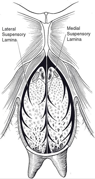
Figure 29-2. Suspensory apparatus of the cow. Udder is shown in transverse section through hindquarters.
practically no vessels or nerves pass through the medial ligament from one half of the udder to the other.
Proximally (close to the body wall), the laminae are thickest. As they descend, they give off sheets of connective tissue that diverge from the midline and interdigitate into the parenchyma of the udder so that the two medial laminae are thinnest near the intermammary groove.The lateral laminae of the suspensory apparatus are composed largely of dense white fibrous connective tissue, making them less elastic than the medial laminae, which are mostly elastic connective tissue. The cranial part of the lateral lamina derives from the aponeurotic tissues of the body wall near the external inguinal ring and more caudally from the regions of the pelvic symphysis and prepubic tendon (the tendon of insertion of the m. rectus abdominis). From its origins, the lateral lamina passes ventrad and around the lateral side of each half of the mammary gland, meeting the medial lamina at the cranial and caudal aspects of each half. Like the medial lamina, the lateral lamina is thick close to the body wall and thins progressively ventrally as it gives off sheets of connective tissue into the substance of the gland.
Blood Supply
The blood supply to the udder is primarily through the external pudendal artery (Fig. 293), a branch of the pudendoepigastric trunk. The external pudendal artery passes downward through the inguinal canal in a more or less tortuous manner and divides into cranial and caudal branches that supply the front- and hindquarters on the same side as the artery. A small artery that may be single or paired (as determined by chance) is the ventral perineal artery, which continues from the internal pudendal artery and passes downward from the vulva just deep to the skin on the median line. The perineal artery usually supplies a small amount of blood to the caudal part of both halves of the udder.
The venous drainage from the udder is largely by way of a venous circle at the base of the udder, where it attaches to the abdominal wall. This venous circle is formed from the main veins that drain the udder.
The external pudendal vein of each side receives blood from both the cranial and caudal quarters of the same side. Cranially, each external pudendal vein is continuous with the caudal superficial epigastric vein and caudally with the perineal vein. An anastomosis between the two caudal superficial epigastric veins just at or in front of the udder completes the venous circle. The caudal superficial epigastric vein passes forward in a sagittal plane lateral to the midline on the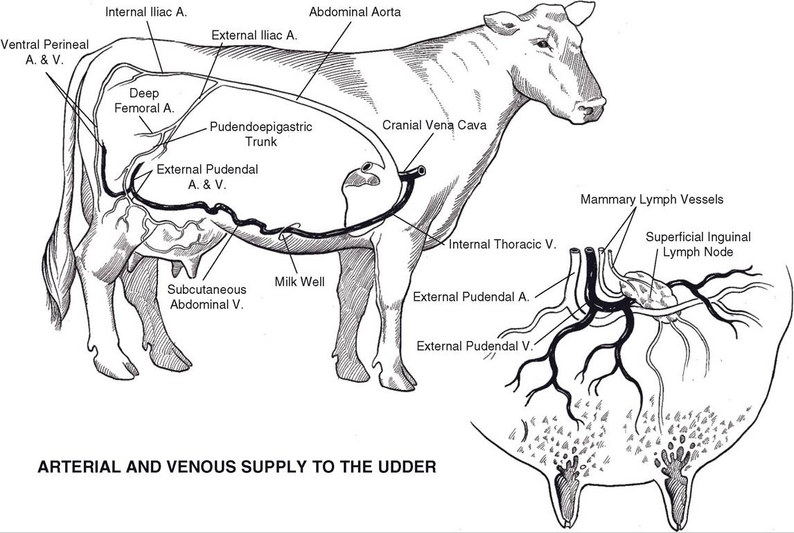
Figure 29-3. Arterial and venous blood supply to the bovine udder. Vessels other than aorta and cranial vena cava are paired.
ventral abdominal wall and joins with the cranial superficial epigastric vein, which ultimately drains to the internal thoracic veins and then to the cranial vena cava. Before the heifer comes into milk, the connection between cranial and caudal superficial epigastrics is poorly developed. During first pregnancy, when the udder undergoes a marked increase in size and, consequently, blood supply, the two veins develop a functional anastomosis, after which they collectively constitute the subcutaneous abdominal vein or the milk vein. In high-producing dairy cows the subcutaneous abdominal vein is large and tortuous. it passes through a foramen in the rectus abdominis muscle (the milk well), joins the internal thoracic vein, and ultimately drains to the cranial vena cava.
Lymphatic Vessels
The lymphatic vessels draining the udder show up rather well superficially just under the skin, particularly in high-producing cattle. They drain from the entire udder, including the teat, to the superficial inguinal (mammary or supramammary) lymph nodes near the superficial (external) inguinal ring above the caudal part of the base of the udder.
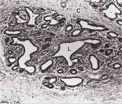
Figure 29-4. Nonlactating mammary gland (cow). A, Gland lobules with inactive alveoli; L, intralobular duct; C, connective tissue. (Reprinted with permission of Wiley-Blackwell from Dellmann, H.D. and Eurell, J. Textbook of Veterinary Histology. 5th ed. Baltimore: Lippincott Williams & Wilkins, 1998.)