Microscopic Anatomy of the Mammary Gland
The mammary gland is classified as a compound tubuloalveolar gland. it consists of a connective tissue interstitium, parenchyma (secretory epithelium), ducts, vessels, and nerves.
in the dry (not lactating) state, the gland has proportionately more stroma than parenchyma (Fig. 29-4); during lactation, the parenchyma undergoes marked growth and constitutes the bulk of the gland (Fig. 29-5).The surface of the bovine and porcine teat is covered with glabrous (hairless), aglandular stratified squamous epithelium. The teats of other domestic species have more typical haired and glandular skin. This is continuous with the stratified squamous epithelium that lines the
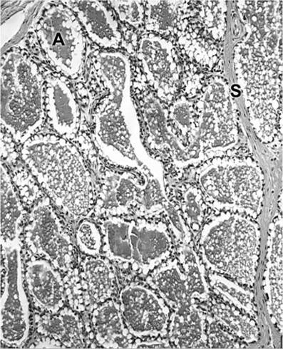
Figure 29-5. Lactating mammary gland. A, alveolus; s, interlobular septum. (Reprinted with permission of Wiley-Blackwell from Eurell, J.A. and Frappier, B.L. DellmannS Textbook of Veterinary Histology, 6th ed. Ames, IA: Blackwell Publishing Professional, 2006.)
papillary duct (streak canal). The duct is encircled by smooth muscle fibers that act as a sphincter.
At the junction of the papillary duct and the teat cistern, the epithelial lining changes abruptly from stratified squamous to stratified columnar epithelium that is usually two cells thick. This stratified columnar epithelium lines the teat and gland cisterns and the larger lactiferous ducts. As the ducts branch and become smaller, the epithelial lining changes first to simple columnar and then to secretory epithelium in the alveoli. The height of the alveolar epithelium varies considerably with the level of activity of the gland.
The mammary gland differs from most other exocrine glands in that the secretory portion is not limited to the terminations of the smallest ducts; milk-secreting structures also empty directly into the larger ducts and directly into the gland cistern and the teat cistern.
A group of alveoli surrounded by a connective tissue septum form a more or less distinct unit called a lobule (Fig.
29-1). A group of lobules within a connective tissue compartment forms a lobe. Correspondingly, the ducts are classified as intralobular, interlobular, intralobar, and interlobar as they increase in size. The alveoli making up the lobule empty into small ducts within the lobule, the intralobular ducts. These intralobular ducts drain into a central collecting space from which the interlobular ducts emerge. Within the lobe, the interlobular ducts unite to form a single intra- lobar duct, which is called an interlobar duct as soon as it emerges from the lobe. The interlobar duct may enter the gland cistern directly, or it may join one or more other interlobar ducts before entering the gland cistern. Many of the ducts have numerous dilations that act, in addition to the lactiferous sinus, as collecting spaces for milk.The alveoli and ducts are surrounded by contractile myoepithelial cells, which are also called basket cells. These cells contract to eject milk (called milk letdown) in response to oxytocin release (discussed later).
in addition to the epithelial parenchyma and the myoepithelial cells, the mammary gland is made up of an interstitium of white fibrous connective tissue and yellow elastic connective tissue. Blood vessels, lymph vessels, and nerves ramify throughout the interstitium to reach the epithelial structures. The veins of the mammary gland are valveless and form a rich network throughout the gland and within the wall of the teat. The vascular layer of the teat is called a corpus cavernosum because of its resemblance to the erectile tissue of the penis in the male. Lymphatic plexuses are found throughout the udder just deep to the skin and scattered throughout the parenchyma of the gland. Nerves appear to be primarily sensory and vasomotor.