Modified Epidermis
Modifications of the epidermis give rise to organs such as hoofs and horns. Many of these tissues have an underlying vascularized connective tissue corium (dermis) that is very prominently folded into papillae or laminae (sheets).
in some places this corium is directly continuous with the underlying periosteum. Because it contains the blood vessels and nerves,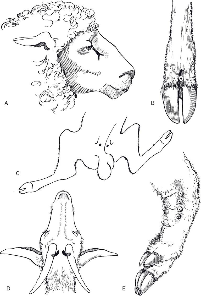
Figure 13-4. Specialized sebaceous glands. A) Infraorbital pouch of the sheep. B) Interdigital pouch of the sheep. C) Inguinal pouch of the sheep. D) Horn glands of the goat. E) Carpal glands of the pig.
the corium is often called the sensitive part of the hoof or horn. The insensitive portions of these structures are derivatives of the overlying epithelium. Nonetheless, it is helpful to keep in mind that the substance of the hoof wall, the horn, and other epidermal modifications is generated by the deepest layer of the epithelium (homologous to the stratum basale of skin) and not by the underlying corium.
Hooves
Hoofed animals are ungulates (L. unguis, nail), and most common farm mammals fall in this category. A defining characteristic of ungulates is the presence of a well-developed hoof associated with the distal phalanx (Fig. 13-5). Although the hoofs of pigs, ruminants, and horses differ significantly in their gross appearance, they share certain features. Like the skin from which they are derived, hoofs have an outer avascular epidermal layer and an inner vascularized dermis; the dermis of hoofs and horns is more commonly called corium.
Different parts of the epidermis and corium of the hoof are named for their location. The outside of the hoof is covered by a thin, waxy layer called the periople. The thick hoof wall grows from a belt of epidermis at the coronary band, the region where haired skin becomes hoof.
The deep side of the hoof wall is intimately connected to the underlying corium, which blends with the periosteum of the distal phalanx. The connection between hoof wall and corium is characterized by interdigitating sheets of hoof wall and corium. These are the laminae, of which there are insensitive laminae (part of the epidermis) and sensitive laminae (part of the corium). The laminae are especially elaborately developed in the equine hoof.The part of the hoof in contact with the ground features a horny sole (extensive in the horse, smaller in other domestic ungulates) and a softer bulb of the hoof. Deep to the bulb of the hoof is a shock-absorbing modification of the subcutis called the digital cushion. The bulb forms a large part of the palmarIplantar aspect of the feet of ruminants and pigs, in which it bears a considerable proportion of the animal’s weight. In contrast, the equine hoof features a keratinized V-shaped frog, which is more flexible than the adjacent sole of the hoof but harder than the bulbs of other ungulates.
The equine hoof is a highly specialized structure and is so important that it will be covered in depth in Chapter 14. The following is a brief overview of the anatomy of the hoofs of artiodactyls, the even-toed ungulates (Fig. 13-6).
A convenient terminology for the digits of even-toed animals is to refer to the digits by number (III and IV in ruminants and pigs), and then relate each digit to the midline of the respective foot. The axial side of the digit is the side closest to the midline of the foot and the abaxial side is the side farthest from the midline of the foot.
The hoof wall consists of a nearly vertical axial portion that reflects sharply caudad at the toe (tip of the hoof) to be continuous with the abaxial portion of the wall. The abaxial hoof wall is convex and the axial wall is concave, and both the axial and abaxial surfaces are continuous with the bulb of the hoof. The lateral digit bears more weight than the medial one (as a clinical consequence, most foot lameness in dairy cattle is referable to the lateral hoof).
In contrast to the horse, the sole and bulb of the foot carry a great deal of weight relative to the walls and toe. The bulb of the porcine hoof is especially prominent, providing a larger proportion of the weight-bearing surface than in ruminants.Horns
Horns of cattle and sheep are formed over the cornual process, a bony core that projects from the frontal bone of the skull (Fig. 13-7). Both male and female cattle of horned breeds have horns, although the female animal’s horns are smaller. In most horned sheep and goat breeds, both males and females have them, although in a few breeds only rams or bucks are horned. Animals that lack horns naturally are polled.
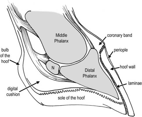
Figure 13-5. Anatomy of the equine hoof. This median section of the hoof illustrates the transition from haired skin to hoof at the coronary band and the relationship between the horny parts of the hoof and the underlying bony structures. (Reprinted with permission of Wiley-Blackwell from Stashak T.S. Adams’ Lameness in Horses. 5th ed. Baltimore: Lippincott Williams & Wilkins, 2002.)
Figure 13-6. Palmar view of the feet of the pig (left) and the ox (right). The accessory digits (dewclaws) of the pig are well developed relative to those of the ox. The bulb of the porcine hoof also makes up a larger portion of the bottom of the foot. Surfaces adjacent to the midline are described as axial, whereas those toward the outside of the hoof are abaxial.
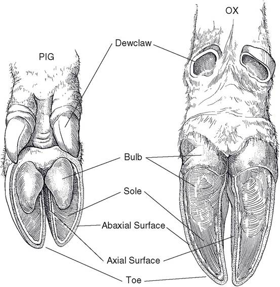
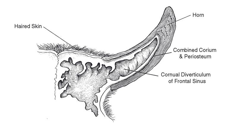
Figure 13-7. Longitudinal section of a horn. The horn is supported by a bony core, the corneal process of the frontal bone, which is invaded by a diverticulum of the frontal sinus. Periosteum and corium are blended on the surface of the cornual process.
The corium of the horn completely envelops the cornual process and blends with its periosteum.
The horn itself consists of dense keratin, much like the hoof wall, and elongates from the base. A soft type of horn called the epikeras covers the surface of the horn at the base and extends a variable distance toward the apex of the horn. The epikeras resembles periople of the hoof.Variations in level of nutrition of the animal are reflected in variations in rapidity of horn growth, resulting in a series of rings on the horn. These alternations in thickness of the horn may reflect seasonal stresses, notably the stress of calving in cows. The age of a cow bearing calves annually may be estimated by counting the rings on the horn.
Dehorning can be accomplished by destroying the corium when only buttons (the small cutaneous primordium of the future horn) are present in the young animal (usually less than 8 weeks for cattle). This is most typically done by surgical removal of the button or by its destruction with a hot iron or with caustic material. For humane reasons, veterinarians administer a local anesthetic block of the cornual nerve before dehorning. After the horn has started to develop, the entire corium and cornual process must be removed along with the horn epidermis and a small amount of adjacent skin to ensure complete dehorning. if any parts are left, an irregular horn stub (a scur) may develop. Dehorning after 3 or 4 months of age risks opening the frontal sinus to the outside, as the cornual diverticulum enlarges into the bony core of the horn as the animal matures.
Dewclaws
The accessory digits, commonly known as dewclaws, of ruminants correspond to digits II (medial) and V (lateral) (Fig. 13-6). Ruminant dewclaws lack well-developed phalanges; their hoofs have a wall and small bulb. Ruminant dewclaws do not bear weight and as a rule have little clinical significance. Dairy farmers occasionally have the medial dewclaws on the pelvic limbs removed as a prophylactic measure against injury to the udder by these horny growths. The dewclaws of pigs, like the weightbearing digits, have three phalanges and a smaller but well-developed hoof. Porcine dewclaws occasionally make contact with the ground when the pig stands on soft surfaces.
Chestnuts and Ergots
Chestnuts are hornlike growths on the medial sides of horses’ limbs. The front chestnuts are proximal to the carpus, and the hind chestnuts are slightly distal to the hocks. The chestnuts are thought to be vestigial metacarpal and metatarsal footpads.
Ergots are small projections of cornified epithelium in the center of the palmar (plantar) part of the fetlock of the horse. The tuft of hair at the fetlock hides the ergot in most instances.