Adnexa of the Skin
Hair
Hair is a defining characteristic of mammals. All common domestic mammals except the pig have abundant hair. There are three main types of hair on domestic mammals: (1) guard hairs, which form the smooth outer coat; (2) wool hairs, also called the undercoat, which are fine and often curly; and (3) tactile hairs, long stiff hairs with specialized innervation that renders them effective as organs of touch.
An individual hair arises from a modification of the epidermis, the hair follicle (Fig. 13-3). The follicle invaginates from the surface of the skin as a double-layered root sheath that surrounds the hair and terminates in a hair bulb of epidermal origin. The hair bulb surrounds a small knob of dermis called the dermal papilla. The internal epithelial root sheath intimately covers the root of the hair and is continuous with the epithelial cells covering the dermal papilla. The external epithelial root sheath surrounds the internal root sheath, is continuous with the epidermis, and gives rise to the sebaceous glands that are associated with hair follicles. The division of the epithelial cells covering the dermal papilla generates the hair itself. Growth and multiplication of these cells extrude the hair from the follicle, causing it to grow.
An individual hair has a medulla at its center, surrounded by a scaly cortex, outside of which is a thin cuticle. All parts of the hair are composed of compressed, keratinized epithelial cells. The bulk of the hair comprises the cortex, which consists of several layers of cornified cells. The amount and type of melanin in cortical cells determine whether the hair will be black, brown, or red. The cuticle is a single layer of thin, clear cells covering the surface of the cortex. The medulla may contain pigment, which has little effect on hair color, but air spaces between medullary cells are believed to
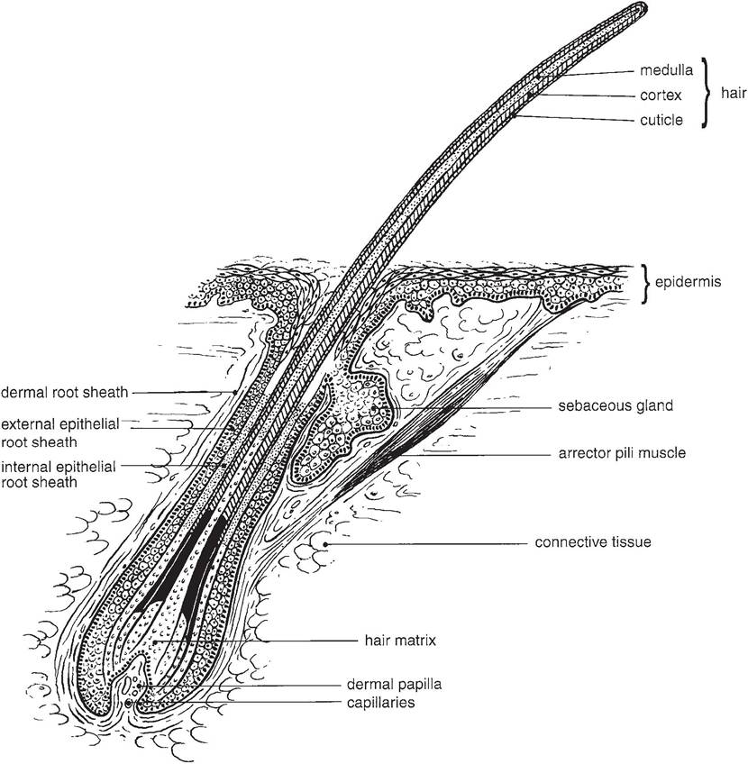
Figure 13-3.
Single hair follicle. Epidermal cells adjacent to the dermal papilla give rise to the keratin of the hair. (Reprinted with permission of Wiley-Blackwell from Eurell, J.A. and Frappier, B.L. DellmannS Textbook of Veterinary Histology, 6th ed. Ames, IA: Blackwell Publishing Professional, 2006.)give a white or silver color to the hair if the cortex lacks pigment. Wool hairs lack a medulla or have only a very small one, accounting for their fine, flexible nature.
Tactile hairs, used as probes or feelers, are also called sinus hairs because a large blood- filled sinus surrounds the deep portions of the follicle. These hairs are thicker and usually longer than guard hairs and are most commonly found on the face, around lips and eyes. These hairs are particularly well supplied with sensory nerve endings that are sensitive to the movement of the hair.
When a hair is ready to shed, the epithelial cells over the papilla stop multiplying and become cornified. The papilla atrophies, and the hair may fall out, be pulled out, or be pushed out by a new hair that develops from epithelial sheath cells in a manner similar to the hair formation just described. A seasonal shedding of the hair coat from the light coat of summer to the heavy coat of winter and back again is characteristic of most domestic species and is largely triggered by changes in the photoperiod.
The arrector pili muscle (plural: arrectores pilorum muscles) is a tiny bundle of smooth muscle fibers that extends from the deep portion of the hair follicle at an angle toward the epidermis (Fig. 13-3). Contraction of the muscle will straighten the hair toward 90°. This orientation increases the insulating properties of the hair coat during exposure to cold and is used by some animals during fight-or-flight reactions, presumably as a means of increasing the apparent size of the animal. The arrectores pilorum muscles are innervated by sympathetic nerves.
Glands
Sebaceous glands are classified as holocrine glands because their oily secretory product, sebum, is produced by disintegration of epithelial cells within the glands.
Most of these oilproducing glands are derived from the external epithelial root sheath and empty their secretion into the hair follicle (Figs. 13-2 and 13-3). Contraction of the arrector pili muscle compresses the glands and aids in emptying them. sebaceous glands that open directly onto the skin surface include those in the ear canal, around the anus, and in the penis, prepuce, and vulva, along with the tarsal glands of the eyelid.Certain animals have specialized sebaceous glands, thought to be marking glands, that are characteristic of their species (Fig. 13-4). Sheep have several cutaneous pouches that are lined with sebaceous glands. These are the (1) infraorbital pouches, found at the medial canthus of the eye and larger in rams than in ewes; (2) interdigital pouches on the midline above the hoofs of all four feet; and (3) inguinal pouches near the base of the udder or scrotum. Goats have sebaceous horn glands caudal to the base of the horn (or where the horn would be in polled animals); secretion in these glands is increased during breeding season and is especially pungent in bucks. in pigs, sebaceous carpal glands are present on the mediopalmar aspect of the carpus in both boars and sows.
Sudoriferous glands or sweat glands (tubular skin glands) can be found over the entire body of farm animals, including the horse, cow, sheep, and pig, although in this last species they are sparse. The horse is the only farm animal that sweats readily. Tubular glands occur on the planum nasolabiale of the cow, planum nasale of the sheep, and planum nasale of the pig (all hairless areas of the nose), where they moisten these surfaces but play little role in cooling. Many modified epithelial structures, including hoofs and horns, lack sweat glands.
Horses are distinguished among domestic animals by their abundant production of sweat. Equine sweat glands, unlike those of most other species, are sensitive to circulating epinephrine, which is why a nervous horse breaks into a sweat in the absence of physical exertion. Equine sweat, moreover, is especially rich in protein; this albuminous sweat will foam when agitated by working muscles. For this reason, a hard-worked horse will lather up on the neck and shoulders and between the pelvic limbs.
The mammary gland is thought to be a modification of tubular sweat glands. its unique importance warrants its own chapter (Chapter 29), and therefore the mammary gland is not considered here.