Motor function in quadrupeds compared to primates
In quadrupedal animals of veterinary interest, the main UMN tracts that influence locomotion originate in the brainstem, whereas the motor cortex/corticospinal system has minimal influence on gait compared with primates and humans.
After experimental removal of the motor cortex of quadrupeds, gait is relatively normal. In contrast to this, damage to the motor cortex in humans can result in marked hemiparesis; this is exemplified by cerebrovascular accidents, or ‘strokes’.Integration of neural functions for locomotion and movement
Basic stepping movements and control systems
Stepping, and the oscillation between extension and flexion of limbs and body, weight bearing and nonweight bearing, is based primarily on reflex circuitry within the spinal cord; it also involves central pattern generators. The flexion of one limb can induce reflex extension in the other limbs, both of the same limb girdle and the other limb girdle (see Fig. 9.1); this utilises the crossed extension reflex. Note that in the normal animal, when recumbent, the crossed extensor reflex is inhibited. In recumbent animals with UMN lesions, active flexion of one limb (e.g. withdrawal reflex) can result in the contralateral limb extending. This indicates loss of descending UMN pathways that inhibit crossed extension reflexes.
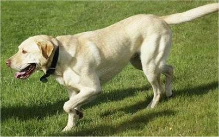
F⅛. 9.1 Extension and flexion of alternate limbs is mediated largely by reflex circuitry within the spinal cord, but supraspinal input is required for locomotion.
The extensor postural thrust reflex is utilised when the foot makes contact with the ground, and the limb joints begin to flex under the effect of gravity. This activates the muscle spindles in extensor muscles, inducing reflex extension and thrust is generated against the ground, resulting in support for the body.
This utilises the same circuitry as the mytotatic reflex.Input, integration and output of the reflexes: Sensory input for the reflexes comes from muscle spindles, Golgi tendon organs, joint and tactile receptors. Integration of that input in the spinal cord causes inhibition or excitation of LMNs, as appropriate, in that same limb, in the opposite limb or the limbs of the other girdle. The activity of the epaxial and hypaxial muscles of the trunk, neck and tail are also interlinked. For example, look at how a cat uses its tail to maintain its balance by offsetting muscle activity in the trunk when it is ‘tightrope walking’ along the top of a narrow fence. Integration of neural activity between the muscles of the limbs and body is done primarily using spinospinal tracts such as the propriospinal tract. This tract connects between spinal cord segments and is located in all funiculi immediately surrounding the grey matter (see Fig. 4.5). Constant proprioceptive input from all body and limb muscles both activates the reflexes and sends sensory information to the cerebellum for use in coordination of motor activity. Proprioceptive input to the forebrain gives rise to conscious awareness of posture and movement; this is called kinaesthesia (see Chapter 6).
The LMNs are stimulated or inhibited by both reflex connections and the input from the UMN system. The UMN tracts stimulate, or inhibit, muscle activity of the limbs and body. In quadrupeds, UMN nuclei of the brain stem are the nuclei primarily responsible for locomotion; there is minimal input from the motor cortex of the cerebrum. Via spinal UMN tracts, UMN nuclei recruit spinal reflex circuitry for locomotion. Their input initiates, regulates, modulates, coordinates and terminates activity in the reflex circuits and in specific LMNs. There are specific locomotive trigger centres in the brainstem. When stimulated, ambulation is triggered. When inhibited, ambulation is terminated.
The UMN tracts primarily responsible for the protraction and support phase are the vestibulospinal and pontine reticulospinal tracts facilitating extensor muscles.
For the retraction/flexor phase, extensor muscle activity must be inhibited and flexor muscle LMNs must be stimulated utilising the medullary reticulospinal and the rubrospinal tracts (Table 4.3). At the end of retraction, extension and subsequently, the support phase, occurs again.Coordination of locomotion
The overall function of the cerebellum is to coordinate agonistic/ antagonistic muscle activity to permit posture and to create movement that occurs at the correct rate, range and force (see Chapter 7).
1. Posture. The cerebellum has an important role in coordinating overall posture.
As described previously, increased load on individual muscles stretches muscle spindles causing reflex contraction of postural muscles, mediated through local spinal circuits. But posture involves the contraction-relaxation of many muscles around numerous joints and that requires overall coordination. The cerebellum’s function is to coordinate the contraction-relaxation of all muscles in the body used for maintaining posture both at rest and during movement.
The postural platform. The cerebellum has a critical role in establishing the postural platform (see Chapter 7). Failure to do so prevents normal, coordinated movement.
2. During movement.
The cerebellum coordinates the initiation of movement, the actual movement itself and the termination of movement; it cannot initiate movement per se. Throughout movement, proprioceptive input from the head, body and limbs, continually informs the cerebellum how much movement has occurred, how fast it is occurring and how forceful the movement is. The cerebellum compares the achieved movement with the planning information it received about that movement. Based on constant proprioceptive feedback, it determines whether the movement is being performed adequately with the correct rate and force. It determines when the correct range of movement has been achieved, and thus when the action should be terminated. The output from the cerebellar cortex is inhibitory.
Thus lesions causing loss of cerebellar output often result in increased rate, range and force of movement. This is called hypermetria.Complex motor activity
For voluntary, learned movement (e.g. capturing ‘prey’ as in Fig. 9.2 or pawing, Fig. 9.3) the planning occurs in the executive motor planning areas of the forebrain. The executive centres draw on integration/interpretation areas associated with a variety of sensory receiving areas (e.g. visual cortex and somatosensory cortex), and memory and behaviour centres of the parietal and temporal lobes. A copy of the planned movement is sent to the cerebellum, which then establishes the appropriate postural platforms. The cerebellum feeds back to the motor planning centres informing them that the posture has been established. The executive centres then direct the senior motor system hierarchy (pyramidal and extrapyramidal systems) and so movement is initiated. Throughout movement, the cerebellum, using constant proprioceptive input, monitors the rate, range and force of movement and sends modifying commands to the UMN centres and constant feedback to the motor planning centres as to how the movement is progressing (see Fig. 7.8).
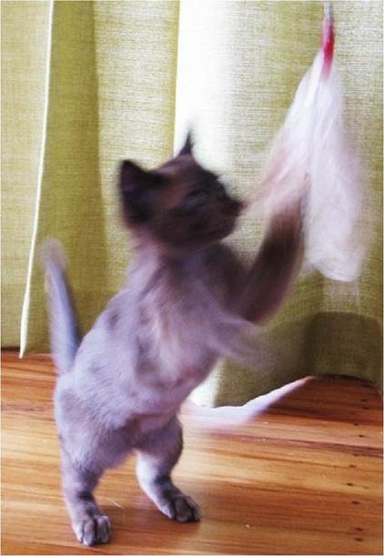
Fig. 9.2 In this fast-moving kitten, the cerebellum has coordinated complex activity in postural muscles to achieve a postural platform that permits use of both forelimbs for a new type of motor function.
Fig. 9.3 Pawing is an example of a complex voluntary movement
(courtesy of Dr. Katherine Houpt, Cornell University).
Repetitive movements and central pattern generators
The CNS contains many neuronal networks that produce oscillatory outputs used for controlling rhythmical motor activity such as locomotion, scratching, chewing, micturition and breathing. Such neural networks are called central pattern generators (CPGs); they contain excitatory and inhibitory neurons.
The CPGs associated with locomotion and scratching have neuronal circuits in the spinal cord intumescences while control centres that initiate and terminate the rhythmical activity are located in the brainstem (Fig. 9.4). Within the spinal cord, the CPG networks are bilaterally symmetrical; there are also interconnections between circuits controlling paired thoracic and pelvic limbs. Spinal cord CPGs can act in isolation from brain and may result in stepping movements; this is called spinal walking and may be seen in animals with chronic spinal cord transection. Spinally generated stepping movements do not produce useful, purposeful locomotion. Input from supraspinal centres in the brain is required for balance, coordination, initiation, regulation, modulation and termination of locomotion.
Fig. 9.4 Rhythmical movements, such as scratching, use spinal cord reflex circuits and are controlled by central pattern generators in the brainstem.
The CPGs associated with the rhythmical movements of the thoracic wall and diaphragm for respiration are located in the brainstem with output to local circuits in the cervical and thoracic spinal cord. The CPG for chewing is also located in the brainstem.
The final common pathway
In summary, influencing the output of the LMN, are incoming peripheral sensory fibres, interneurons from many spinal cord segments, UMN, and, indirectly, the cerebellum and executive motor centres of the forebrain. There is a complex system of checks and balances, whereby the senior and executive components of the motor system facilitate or inhibit the LMN activity. These influences act directly on individual groups of LMNs or indirectly by activating reflex circuits.
The following points are given to illustrate some of the complexities that are used to achieve optimal motor function.
Facilitation of LMNs occurs by output from many areas, including limited areas of the cerebral cortex, the globus pallidus of the basal nuclei, the red, pontine reticular and vestibular nuclei.
Indirect facilitation also occurs by inhibition of inhibitory pathways. For example, the medial medullary reticular formation gives rise to the medullary reticulospinal tract, which exerts a massive inhibitory effect on LMNs supplying extensor muscles. However, the lateral medullary reticular formation can inhibit it, thereby resulting in indirect facilitation of LMN activity.The LMNs are directly inhibited by the medullary reticulospinal tract and are also indirectly inhibited by the executive management which can inhibit certain UMN centres of the brainstem. The cerebral cortex also projects to the substantia nigra of the midbrain, which has an inhibitory effect on the basal nuclei, dampening down the stimulatory effect of the globus pallidus on the UMN nuclei of the brainstem.
Ultimately, the activity of motor system hierarchy is expressed as movement through the activation, or inhibition, of α-LMN firing. Again, this often occurs indirectly as a consequence of γ-LMN-1a (see Chapter 5) activity. Thus, the α-LMN is referred to as the final common pathway. Through the α-LMN, a myriad of inputs is finally expressed. The activity of all these inputs is summated and if facilitation exceeds inhibition, the α-LMN is activated and the muscle will contract at a strength that is proportional to the frequency of firing. This is occurring constantly and in hundreds of muscles around the body. Simultaneously, there are changes in the activity of muscles antagonising the contracting muscles. If the antagonist muscle(s) is inhibited, agonist contraction results in joint movement. If the antagonist muscle(s) also contracts, then the joint position is fixed. Isometric contraction of agonists and antagonists and fixing of joint position, is the basis of static posture. The contraction-relaxation of so many muscles is coordinated and regulated by the organising centre, the cerebellum.