MUSCLES CONTROLLING MOVEMENTS OF THE HEAD
The four straight and two oblique muscles associated with the atlantooccipital and atlantoaxial joints form a group of their own.
The rectus capitis dorsalis major (Figure 12-11/2) arises from the spine of the axis, just cranial to the attachment of the nuchal ligament, and inserts on the nuchal surface of the skull, ventral to the insertion of the semispinalis capitis, by which it is covered.
The rectus capitis dorsalis minor (Figure 12-11∕to the cervical vertebrae is indicated for vertebral fractures; in this approach the biventer cervicis and the rectus capitis dorsalis major are exposed cranially, and the nuchal ligament, the spinalis et semispinalis cervicis, and multifidus cervicis muscles are exposed more caudally. The vertebral artery lies in the rectus capitis dorsalis major, ventrolateral to the synovial joint C1∕C2 and must be avoided as the dissection is continued laterally.
The dorsal approach to the caudal cervical and cranial thoracic vertebrae for dorsal laminectomy (removal of part of the vertebral arch) and fracture
Figure 12-12
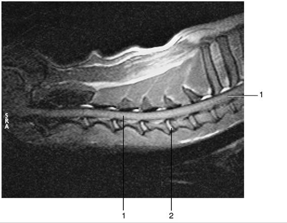
Midsagittal section of cervical region of dog, T2 weighted. 1, Spinal cord; 2, nucleus pulposus.
repair first exposes the aponeuroses of the trapezius cranially and the rhomboid caudally. Then the subscapulares, splenius, and serratus dorsalis are exposed by lateral retraction of the trapezius and rhomboid muscles and the scapula. Finally, the semispinalis capitis, longissimus cervicis, the nuchal ligament, and the dorsal spines of the vertebrae are exposed by lateral retraction of the splenius and serratus dorsalis. The deep cervical artery passes through the semispinalis capitis.
A dorsal approach to the thoracolumbar vertebrae is indicated for dorsal laminectomy and thoracolumbar fractures. Lateral retraction of the lumbar fascia exposes the longissimus lumborum and multifidi caudally and the spinalis et semispinalis thoracis cranially. The multifidus, interspinalis, and rotatores longi are elevated from the spinous processes and vertebral arches. The dorsal branch of each spinal nerve emerges just cranial and ventral to the insertions of the longissimus on the accessory processes.