Muscles of the Trunk and Neck
Extensors of the Vertebral Column
The group of muscles dorsal to the transverse processes of the vertebrae on either side of the spinous processes are the epaxial muscles. These make up the loin muscles and continue forward to the head.
Collectively, they are referred to as the m. erector spinae. In domestic animals, the largest of these muscles is known as the m. longissimus. It is composed of many small bundles of muscle fibers that extend from vertebral transverse processes to spinous processes, from transverse processes to transverse processes, or between spinous processes. As these attachments may extend from one vertebra to the next or overlap one or more vertebrae, there are many possibilities for naming individual muscles and a variety of individual muscle actions. As a general description, however, we can say that these muscles are responsible for extension, and when acting unilaterally, lateral flexion of the vertebral column. They may also cause slight rotation (twisting) of the vertebral column, as seen when a bucking horse throws the front feet to one side and hind feet to the opposite side.other epaxial muscle groups include the medial transversospinalis system, whose fibers span one or more vertebrae from transverse to spinous processes, and the iliocostalis system. The latter is the most lateral group of the epaxial muscles (Fig. 7-12).
The same general arrangement of muscles is continued into the neck, where much greater flexibility is evidenced. The dorsal neck muscles, which extend (raise) the head and neck, are well developed to support the head. The large extensor muscles of the head originate from the vertebrae in the region of the withers and insert on the occipital bone of the skull. The most superficial of these muscles (other than the m. trapezius, which does not originate from the vertebrae) is the m.
splenius, and deep to it is the m. complexus (Figs. 7-1 to 7-3). Other muscles that actively extend the head and neck include the m. rhomboideus, continuations of the m. longissimus, and the rectus capitis and obliquus capitis groups. Deeper muscles that extend from one vertebra to the next also aid in movements of the neck. in addition to these muscles, a heavy elastic band, the ligamentum nuchae (nuchal ligament), reaches from the withers to the skull. The ligamentum nuchae gives considerable aid to the muscles that extend and support the head and neck.Flexors of the Vertebral Column
Muscles ventral to the transverse processes of the vertebrae are the hypaxial muscles. They tend to flex the trunk, neck, and head. These include the m. sternocephalicus, which extends from the sternum to the mandible in the horse and to the mandible and mastoid process of the skull in ruminants. in addition, the m. sternothyroideus, m. sternohyoideus, m. longus colli, and m. longus capitis are flexors.
Abdominal Muscles
The muscles that form the bulk of the abdominal wall support the organs of digestion and many of the reproductive organs, particularly the gravid (pregnant) uterus. The abdominal muscles may act to flex the vertebral column. if contracting on one side only, they flex it laterally or even twist the vertebral column. These muscles are important in emptying the contents of the digestive tract (defecation), urinary tract (urination, also called micturition), and female reproductive tract at birthing (parturition). The abdominal muscles are used in regurgitation and vomiting and serve as strong muscles for forced expiration of air from the lungs, as seen during coughing or sneezing.
The abdominal muscles are arranged in layers much like plywood, with the muscle fibers running in different directions. Most of these muscles have broad aponeurotic insertions that meet at the midventral line known as the linea alba (white line).
The external abdominal oblique m.
(m. obliquus externus abdominis) is the most superficial. The fibers of this muscle lie obliquely ventrad and caudad. its origin is from the last few ribs and thoracolumbar (lumbodor- sal) fascia over the back and loins. The insertion is by means of a broad flat tendon (aponeurosis) that meets the insertion of the muscle from the opposite side at the linea alba. Caudally, the muscle is continued by an aponeurosis, sometimes called the inguinal ligament, at the junction of abdominal wall to pelvic limb. This ligament forms the superficial wall of the inguinal canal for the passage of the spermatic cord of the male. it contains a slit, the superficial inguinal ring, through which the spermatic cord passes from the inguinal canal into the scrotum.The internal abdominal oblique m. (m. obliquus internus abdominis) is immediately deep to the external abdominal oblique muscle. its fibers pass obliquely ventrad and craniad, and the muscle also inserts on the linea alba by means of an aponeurosis. in some animals, this muscle forms the deep wall of the inguinal canal and also the deep inguinal ring. The most
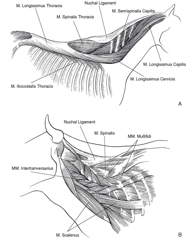
Figure 7-12. Epaxial muscles. A) Superficial muscles of the horse. B) Deeper muscles of the ox.
caudal group of fibers from the internal abdominal oblique muscle passes through the inguinal canal with the spermatic cord and attaches to the outer covering of the testicle. This slip of muscle constitutes the cremaster m., which pulls the testicle toward the inguinal canal. In some animals, such as rodents, the testicle is retracted into the abdominal cavity except during breeding seasons.
The m. transversus abdominis is the deepest of the abdominal muscles. It originates from the deepest layer of thoracolumbar fascia, and the fibers are directed perpendicular to the long axis of the body to insert on the linea alba.
The m. rectus abdominis forms the muscular floor of the abdomen.
It originates from the cartilages of the ribs and the sternum. The fibers run directly caudad in a horizontal plane to attach to the pubis by means of a strong prepubic tendon. The m. rectus abdominis characteristically is divided by a series of tendinous intersections.Muscles of Respiration
The muscles of respiration are either expiratory, forcing air out of the lungs by decreasing the size of the thorax, or inspiratory, causing air to enter the lungs by increasing the size of the thorax.
The diaphragm is the chief muscle of inspiration. it is a dome-shaped sheet of muscle separating the thoracic and abdominal cavities. it projects into the thorax. Contraction of the fibers of the diaphragm tends to flatten the diaphragm and force the abdominal viscera caudad, further into the abdomen. This, in effect, increases the volume of the thorax and lowers intrathoracic pressure, drawing air into the lungs.
The external intercostal mm. (mm. intercostales externi) extend from each rib to the next rib behind. The fibers are directed ventrad and caudad similarly to the external abdominal oblique muscle. When these muscles contract, they tend to rotate the ribs upward and forward, increasing the size of the thorax. The internal intercostal mm. (mm. intercostales interni), which lie deep to the external intercostal muscles, run from each rib to the next one in front, and the fibers are directed ventrad and craniad. Although not all studies are in agreement, most anatomists describe their action as reducing the volume of the thorax and therefore aiding in forced expiration. some authorities believe that both the external and internal intercostal muscles may function in both inspiration and expiration.
As mentioned previously, the abdominal muscles may act as muscles of expiration by forcing the abdominal viscera against the diaphragm, decreasing the size of the thorax.