Neurons Have Four Distinct Anatomical Regions
A typical neuron has four morphologically defined regions (Figure 4-1): the dendrites, the cell body, the axon, and the presynaptic terminals of the axon. These four anatomical regions are important in the major electrical and chemical responsibilities of neurons: receiving signals from the presynaptic terminals of other neurons (on dendrites), integrating these often-opposing signals (on the initial segment of the axon), transmitting action potential impulses along the axon, and signaling an adjacent cell from the presynaptic terminal.
These functions are collectively analogous to the general role of the nervous system: collecting information from the environment, integrating that information, and producing an output that can change the environment.The cell body (also called the soma or perikaryon) plays a critical role in manufacturing proteins essential for neuronal function. Four organelles are particularly important for this purpose: the nucleus, containing the blueprint for protein assembly; the free ribosomes, which assemble cytosolic proteins; the rough endoplasmic reticulum, in which secretory and membrane proteins are assembled; and the Golgi apparatus, which further processes and sorts secretory and membrane components for transport. The cell body usually gives rise to several branchlike extensions, called dendrites, whose surface area and extent greatly exceed those
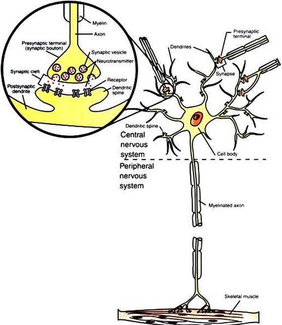
FIGURE 4-1 A typical neuron has four functionally important regions.The cell body manufactures proteins to maintain the neuron; the dendrites receive signals from neighboring neurons; the axon integrates these signals and transmits action potentials some distance along the cell; and the presynaptic terminal signals adjacent cells.The inset shows an enlargement of the circled synapse.
of the cell body. The dendrites serve as the major receptive apparatus of the neuron, receiving signals from neighboring neurons. These signals affect specialized receptor proteins (receptors) that reside on the dendrites. The cell body also gives rise to the axon, a tubular process that is often long (>1 meter in some large animals). The axon is the conducting unit of the neuron, rapidly transmitting an electrical impulse (the action potential) from its initial segment at the cell body to the other end of the axon at the presynaptic terminal. Axons lack ribosomes and therefore cannot synthesize proteins. Instead, macromolecules are synthesized in the cell body and are carried along the axon to distant axonal regions and to the presynaptic terminals by a process called axoplasmic transport. Large axons are surrounded by a fatty, insulating coating called myelin. In the peripheral nervous system, myelin is formed by Schwann cells, specialized glial cells that wrap around the axon much like toilet paper wrapped around a broomstick. A similar function is performed by glial cells called oligodendrocytes in the central nervous system. The myelin sheath is interrupted at regular intervals by spaces called nodes of Ranvier. The myelin sheath significantly increases the speed of action potential conduction along the axon.
Axons branch near their ends into several specialized endings called presynaptic terminals (or “synaptic boutons”). When the action potential rapidly arrives, these presynaptic terminals transmit a chemical signal to an adjacent cell, usually another neuron or a muscle cell. The site of contact of the presynaptic terminal with the adjacent cell is called the synapse, shown in the inset in Figure 4-1. It is formed by the presynaptic terminal of one cell (presynaptic cell), the receptive surface of the adjacent cell (postsynaptic cell), and the space between these two cells (the synaptic cleft). Presynaptic terminals contain chemical transmitter-filled synaptic vesicles that can release their contents into the synaptic cleft. The presynaptic terminals of an axon usually contact the receptive surface of an adjacent neuron or muscle cell, usually on the neuron’s dendrites, but sometimes this contact is made on the cell body or, occasionally, on the presynaptic terminals of another cell (e.g., for presynaptic inhibition). On many neurons, presynaptic terminals often synapse on small protrusions of the dendritic membrane called dendritic spines. The receptive surface of the postsynaptic cell contains specialized receptors for the chemical transmitter released from the presynaptic terminal.