Ossification
Ossification is the formation of true bone by deposition of calcium salts in a matrix of osteoid tissue. Calcification refers to the deposition of calcium salts in any tissue. Calcification of tissue other than osteoid is usually associated with some pathologic process.
Regardless of the location, the sequence of actual bone formation consists of osteoblasts laying down osteoid tissue that is subsequently calcified under the influence of the enzyme alkaline phosphatase. A local area of bone formation is called a center of ossification (Fig. 5-5). The environment in which bone forms determines whether the type of ossification is endochondral or intramembranous.
Endochondral (Intracartilaginous) Ossification
During fetal development, most of the skeleton first develops as a cartilage pattern or model, and then the cartilage of this model is gradually replaced by bone. This process is called endochondral ossification. The center of ossification that develops in the midshaft region of a long bone is the primary ossification center (Fig. 5-5). Secondary ossification centers then develop near the ends of long bones. These ossification centers grow and expand, but a region of cartilage, the physis, still separates the centers during growth and development. Chondrocytes within this region continue to proliferate and produce cartilage to provide this separation and thus allow for continued growth in the length of the long bone.
The midshaft region of a long bone that contains the primary ossification center is the diaphysis; each end that contains a secondary ossification center is an epiphysis (Fig. 5-5). As animals grow and mature, the region of cartilage that separates the bony diaphysis and epiphyses continues to narrow. The epiphyseal plate, another term used to describe this region of cartilage in growing animals, emphasizes how narrow it may become.
When the cartilage in the epiphyseal plates is completely replaced by bone, increases in length, hence growth in stature of the animal, is impossible. This is epiphyseal closure. An epiphyseal line can often be seen on bones where this closure has occurred. The region of a long bone where the diaphysis and an epiphysis meet is a metaphysis (Fig. 5-5).
several hormones affect the rate of growth of long bones, but growth hormone and sex hormones (androgens and estrogens) are key regulators. in general, growth hormone promotes elongation of long bones, and the sex hormones promote growth and epiphyseal closure. Growth hormone itself has little direct effect on chondrocytes within epiphyseal plates. it stimulates other cells within the area of the plates and in the liver to produce peptides, insulin-like growth factors (IGFs), which in turn stimulate the chondrocytes to proliferate and increase their rate of cartilage production. This provides more cartilage in which bone can form to increase the length of the long bone. The critical role of IGFs has been confirmed in some human dwarfism and in African Pygmies who have normal blood levels of growth hormone but who do not grow tall due to of low levels of IGFs.
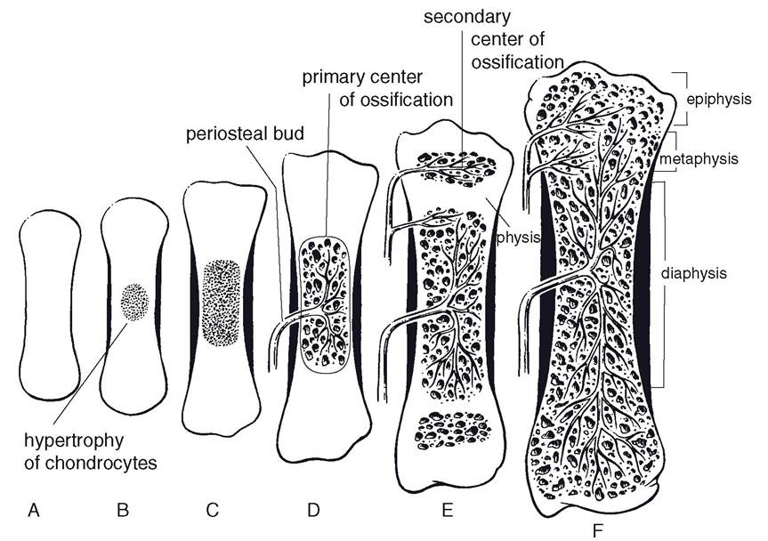
Figure 5-5. The stages of endochondral ossification of a long bone. A) A cartilage model forms initially. B) The chondrocytes in the center of the model hypertrophy. C) A bony collar begins to form around the cartilage model. D) Blood vessels from the periosteum (periosteal bud) invade the cartilage model, bringing bone-forming cells to initiate the primary center of ossification. E) The physis and secondary centers of ossification are established. F) The growth plate closes in the mature bone, and a confluent marrow cavity from the epiphysis to the diaphysis is formed. (Reprinted with permission of Wiley-Blackwell from Dellmann, H.D. and Eurell, J.
Textbook of Veterinary Histology. 5th ed. Baltimore: Lippincott Williams & Wilkins, 1998.)Androgens, such as the sex hormone testosterone, and estrogens have a variety of complex effects on the rates of bone growth. The well- recognized growth spurt associated with puberty is thought to be due to stimulatory effects of androgens and estrogens that increase in circulation at this time. Androgens appear to have a greater stimulatory effect than estrogens, and this difference is responsible, in part, for differences in body size between males and females. The stimulatory effects of androgens on growth are in part due to their ability to increase the secretion of growth hormone. While sex hormones are capable of stimulating the rate of growth, they also bring about epiphyseal closure, which ultimately limits body size. The mechanisms responsible for this effect are not completely understood, but differences in the magnitude of their stimulatory effect on ossification versus their stimulatory effect on cartilage production may be responsible.
Intramembranous Ossification
Many of the flat bones, such as bones of the skull, are preformed in a fibrous membrane, or matrix, which is infiltrated with osteoid tissue. The osteoid tissue calcifies to form true bone. The layers of periosteum on either side of the bone then form additional bone. Like long bones of the limbs, large flat bones in mature animals consist of compact bone surrounding a cancellous bone core.