Microscopic Anatomy and Formation of Bone
About a third of the weight of bone consists of an organic framework of fibrous tissue and cells. This organic matter mainly consists of collagen and polysaccharides called glycosaminoglycans (GAGs), which contain chondroitin sulfate.
They give resilience and toughness to bones. The remaining two-thirds of bone weight consists of inorganic calcium and phosphorus salts in the organic framework. About 80% of these salts are calcium phosphate, and the remainder is primarily calcium carbonate and magnesium phosphate. The calcium phosphate is primarily found in hydroxyapatite crystals formed with calcium hydroxide. These salts give hardness and rigidity to bones and make them resist the passage of x-rays. If the inorganic salts are removed by soaking a bone in dilute acid, the resulting decalcified bone will retain its original form but will be flexible enough to be tied in a knot. On the other hand, if the organic matter is removed by charring in a furnace so that only the inorganic salts remain, the bone will retain its form but be brittle and break unless handled with extreme care.Mature bone consists of osteocytes (bone cells) surrounded by an intercellular matrix composed of calcified osteoid material. The
osteocytes are in small cavities in the bone called lacunae (meaning little lakes) (Fig. 5-1). A system of tiny canals called canaliculi connects the lacunae within the substance of the bone. Even though bone is highly vascular, with capillaries close together, the canaliculi transmit tissue fluid that is essential for maintaining the life of the osteocytes.
Both the lacunae and canaliculi are formed because cytoplasmic processes connect the osteoblasts (bone-forming cells) at the time the osteoid material is laid down. Thus the cells and their processes act as a mold until the osteoid tissue is set and mineralized.
The cytoplasm is then partially withdrawn, leaving the cells, now known as osteocytes, in the lacunae, which are connected by canaliculi containing cytoplasmic extensions.Cancellous bone, or spongy bone, consists of a network of fingerlike bony spicules, or trabeculae. This type of bone is found in the
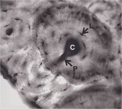
Figure 5-1. Unstained ground bone. Osteocytes in lacunae (L) and fine canaliculi extend from each lacuna. The central (C) and perforating (P) canals contain blood vessels, nerves, and lymphatics. (Reprinted with permission of Wiley-Blackwell from Dellmann, H.D. and Eurell, J. Textbook of Veterinary Histology. 5th ed. Baltimore: Lippincott Williams & Wilkins, 1998.)
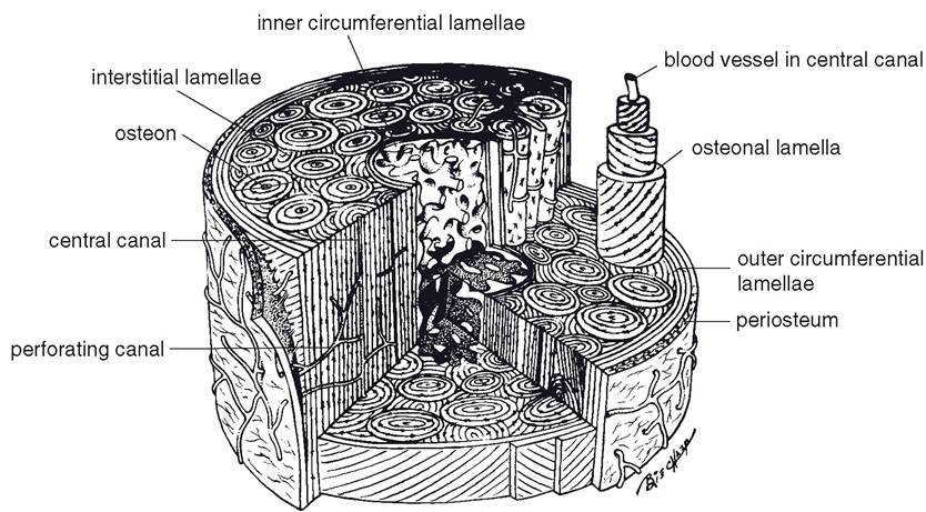
Figure 5-2. The structural unit of compact bone is the osteon. An osteon is telescoped to show the concentric layers of bone that surround a central canal. interstitial lamellae of bone fill the space between osteons, and the inner and outer surfaces are formed by inner and outer circumferential lamellae. (Reprinted with permission of Wiley-Blackwell from Dellmann, H.D. and Eurell, J. Textbook of Veterinary Histology. 5th ed. Baltimore: Lippincott Williams & Wilkins, 1998.)
extremities of long bones, where resistance to compression without excessive weight is needed. Flat bones between two layers of compact bone, as in the skull, are also cancellous. The spicules of bone are arranged so as to resist stresses and strains imposed on the bone by weight or pull of muscles.
compact bone, found in the shafts of long bones, consists primarily of many laminated tubes known as osteonal systems (formally termed haversian systems). Each osteon consists of one central canal containing vessels and nerves surrounded by circular plates of bone (osteonal lamellae) forming the laminated cylinder (Fig.
5-2). These plates are laid down in a centripetal fashion (from the periphery toward the center). After the bone is formed, the osteoblasts that became embedded in the bone substance are called osteocytes. in general these osteons are added on the periphery of the shaft of a bone as the bone increases in diameter. Blood vessels extend from the periosteum to central canals through perforating canals (also known as Volkmann’s canals), which often travel at right angles to the central canals (Fig. 5-2).osteoblasts usually come from mesenchymal cells, the parent cells of all connective tissues. The osteoblasts divide readily, but only a portion of the new cells actually secretes osteoid substance and forms bone; the rest is held in reserve as the osteogenic layer of the periosteum and endosteum within the marrow cavity and central canals. These reserve cells divide and form more osteoblasts whenever more bone is needed, as in repair of fractures, response to stress, or growth. Because the intercellular matrix is unyielding, bone can be added only on the surface, and the osteocytes (mature osteoblasts) probably have lost the ability to divide.
As the shaft of a long bone enlarges in diameter as a result of the activity of the osteogenic layer of the periosteum, bone along the inner surface is normally resorbed to increase the size of the marrow cavity (Fig. 5-3). Resorption of bone may also occur under abnormal conditions, such as during a period of calcium defi-
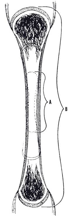
Figure 5-3. Remodeling that occurs as a long bone increases in size. Both resorption and deposition of bone take place. A, Size and shape of young bone; B, size and shape of mature bone.
ficiency. Whenever bone is resorbed (under normal or abnormal conditions), large multinucleated cells called osteoclasts (bonedestroying cells) are usually found (Fig. 5-4). These cells, derived from macrophages, take an active part in bone destruction by releasing organic acids and enzymes.
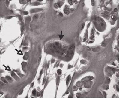
Figure 5-4. Osteoclast (solid arrow) resorbs bone. Osteoblasts (open arrows) form bone. (Reprinted with permission of Wiley-Blackwell from Dellmann, H.D. and Eurell, J. Textbook of Veterinary Histology. 5th ed. Baltimore: Lippincott Williams & Wilkins, 1998.)